From benchtop to bedside:

3D Bioprinting Cartilage to Develop Treatments for Knee Injuries
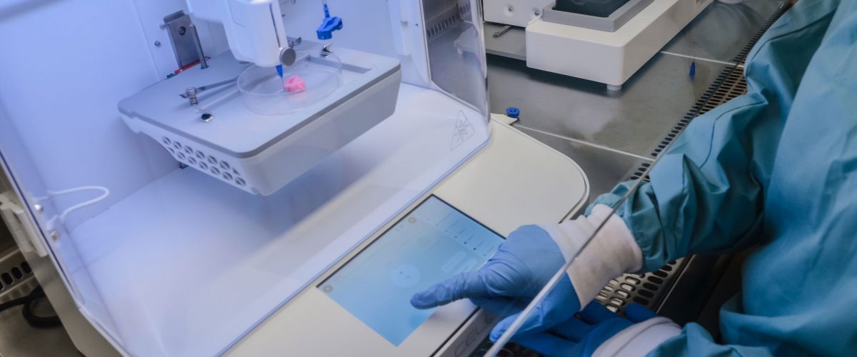
Stina-Customer-Spotlight-Using-the-BIO-X

Translating stem cell research into actionable therapies to improve life quality

Personalized medicine has been on the forefront of many researchers’ minds. An elusive target to develop therapies that treat each patient for exactly their ailments that many spend their time and energy on. Stina Simonsson, a professor and researcher out of Gothenburg University leads a team focused on such work. The group has developed an in-depth understanding of stem cells, and how these wonderous building blocks can be reprogrammed to create life changing therapies.

Developing treatments for knee injuries

As it stands today, one out of four Swedes over the age of 45 suffer from some degree of osteoarthritis. Dr. Simonsson and her group began their research to develop a solution for this widespread problem by developing mini-knees in test tubes, culturing iPSC’s and having them differentiate into cartilage cells. These mini-knees helped pave the way to understand disease mechanisms and what causes the cartilage to decay.

Hear what Stina Simonsson has to say

Bioprinting personalized iPS cell-laden implants for a perfect fit

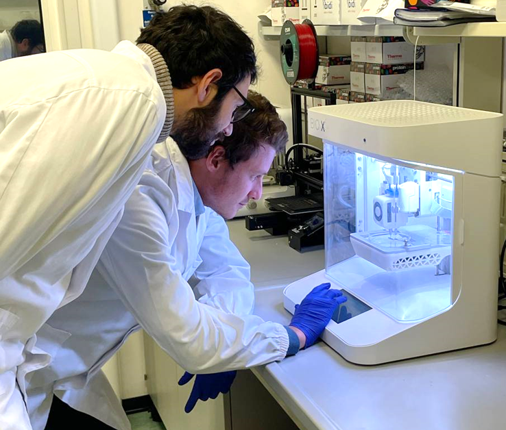
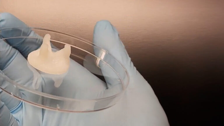
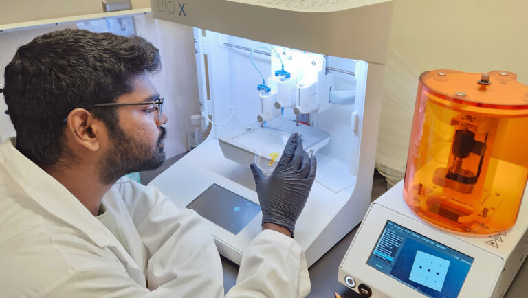
Armed with an understanding of what causes stem cells to differentiate into cartilage, Dr. Simonsson and her team unlocked a new degree of personalization by taking patient scans and using the BIO X to develop patient specific constructs. These iPSC laden constructs were printed using a combination Nanocellulose and alginate similar to CELLINK’s bioink. Optimizing the bioink allowed for a high degree of fidelity for the print, but more importantly, safety during the printing process, which in turn ensured high cell viability and high probability of differentiation. This process entailed a robust construct with ECM proteins like Collagen Type II and GAGS being developed in abundance. This bioprinted cartilage when compared to in vivo cartilage was indiscernible, even by trained surgical eyes.

Taking on demand cartilage from basic research to clinical therapies

Having successfully demonstrated that on demand cartilage can be bioprinted, the team now turns their eyes to translating this research into clinical therapies. To do so, the team is faced with a number of challenges. One such challenge is finding a suitable material that possesses the same rheological properties as their bioink but has a structure even closer to humans and also enables a simpler path through the regulatory obstacles that must be navigated as research is turned into treatments.
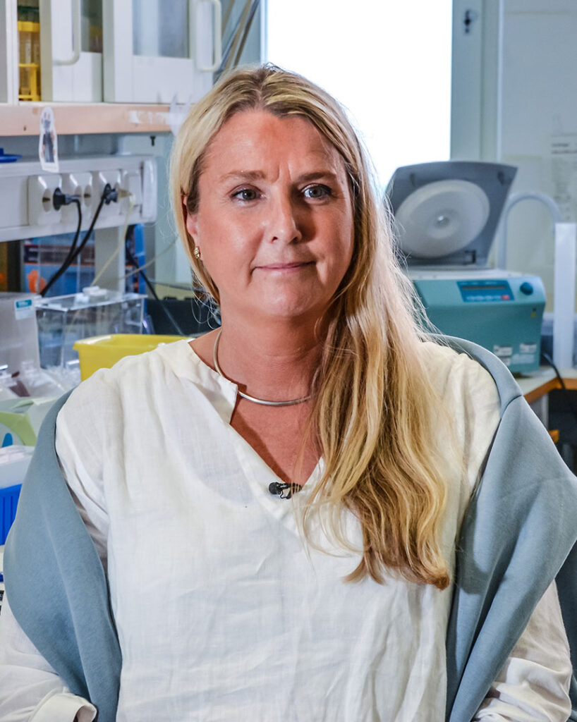
Dr. Stina Simonsson at Gothenburg University
“CELLINK technology and bioinks has helped me tremendously in my research and I wouldn’t have gotten so far in my research without their support,” says Stina Simonsson.

More Customer Spotlights

Bioprinted Skeletal Muscle Tissue with a Perfusable Microchannel Network

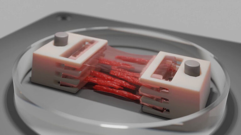
Skeletal muscle tissue (SMT), the body’s largest organ by mass, plays a vital role in movement, posture, breathing, overall physiology and energy homeostasis. However, genetic disorders, diseases, injuries, and aging can impair this essential organ, significantly impacting health and quality of life. Traditional 3D culture systems have struggled to replicate the complex structure and functionality of native muscle tissue, particularly at larger, centimeter scales. Using advanced bioprinting technologies, Dr. Miriam Filippi and her group from the Soft Robotics Laboratory (led by Prof. Robert Katzschmann at ETH Zurich) have developed biohybrid SMT constructs that effectively mimic native muscle tissue.

Innovative Silk Bioink for 3D Bioprinted Bone Marrow Tissue Models

There is a rising need of platelet units, which is primarily sourced from healthy donors. However, the natural production of platelets coupled with the brief shelf life does not meet the demand by the healthcare industry. Prof. Alessandra Balduini and her team at the University of Pavia are taking this challenge head-on by initiating the EIC Transition SILKink project, which aims to produce a groundbreaking platform that combines the use of natural silk and 3D bioprinting to recreate the environment of the human bone marrow where platelets are produced.

Bioprinting pediatric heart valves that stand the test of time

Heart valve disease is a significant global health issue, affecting millions of people and contributing to a substantial number of cardiovascular-related deaths. Traditional mechanical replacements, while life-saving, present several challenges, especially over time. Most mechanical valves do not grow or adapt with the patient’s body, leading to the need for multiple invasive surgeries. This not only poses inherent risks but also significantly impacts the patient’s quality of life. Issues such as blood clots, calcification, and severe inflammation further complicate the situation. These challenges become even more critical in pediatric cases, where conventional solutions, while still life-saving, may reduce life expectancy by up to 50%. Professor Savoji, Arman Jafari, a PhD student in his lab, and a dedicated team at the University of Montreal have employed bioprinting and their BIO X6 to address these challenges and develop a method to bioprint heart valve replacements.

Empowering Highly Motivated Researchers

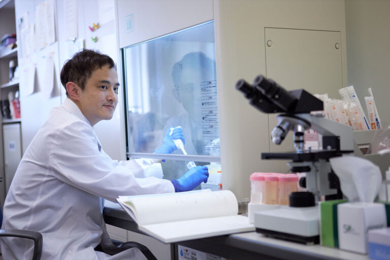
We have interviewed Takaaki Arahira, the Associate Professor of the Department of Information Networks, Faculty of Management and Information Sciences at Kyushu Institute of Information Sciences.