A Colorectal Cancer 3D Bioprinting Platform for Disease Modeling

Chief Assistant Professor Yordan Sbirkov, PhD, and Professor Victoria Sarafian, MD, PhD, DSc, work at the Medical University of Plovdiv in Bulgaria, where Dr. Sarafian is head of the Department of Medical Biology. They have both collaborated over the years with Professor Colin McGuckin, PhD, founder and chief scientific officer at CTIBiotech in Lyon, France, including on a 2021 publication “A Colorectal Cancer 3D Bioprinting Workflow as a Platform for Disease Modeling and Chemotherapeutic Screening.” During a recent intercontinental video conference, they discussed developing their original protocol, collaborating with CELLINK, and the singular challenges of working with tumor samples from bacteria-filled human guts.

Tell us about your colorectal cancer research

Yordan Sbirkov
In our paper, we tested whether a 3D bioprinted model of colorectal cancer better represents patient tumors compared to standard monolayer or 2D cell culture methods. In our comparisons, we saw differences in gene expression, morphology, growth patterns, even drug responses. Overall, our 3D model was more drug resistant than the 2D cell culture control.
Colin McGuckin
Sometimes researchers screen a chemotherapy drug on 2D cancer cells, and it works very well. But when the same drug is used on the patient, it doesn’t work at all. And that is one of the reasons why so many drugs fail in clinical trials. By 3D bioprinting a tumor model for preclinical drug screening, we can better work out if the chemotherapy is getting into the cancer cells in a more relevant 3D model of the in vivo microenvironment. Then we can test new strategies to penetrate the chemotherapy into the part of the tumor that needs to be killed.
Victoria Sarafian
Colorectal cancer research was not a new topic for our department at the Medical University of Plovdiv. But using 3D bioprinting to develop a drug testing platform for personalized treatments was new and exciting. When we started the project some 3 years ago, we were enthusiastic to find only four papers published on 3D bioprinted colorectal cancer models. As we set about developing our protocol, we had to deal with contamination with microbiota and many other challenges. That might explain why there weren’t many papers on the topic.

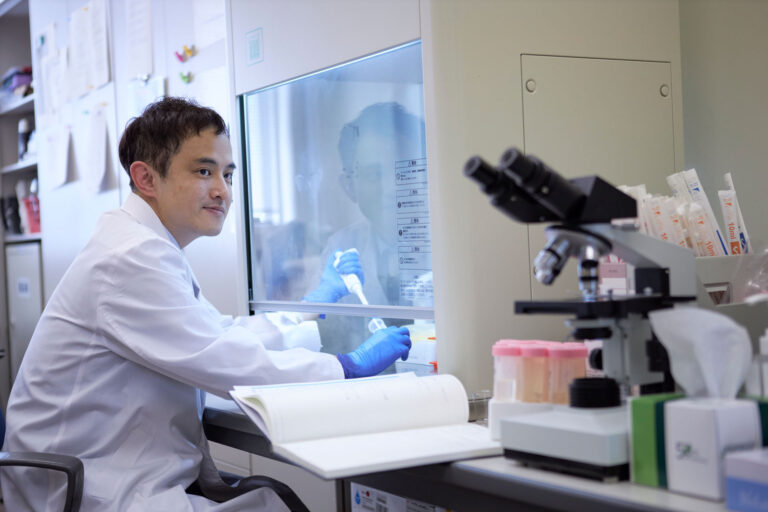
“The BIO X’s flexibility, speed and reproducibility were instrumental in successfully printing our 3D colorectal cancer models,” says Dr. Sbirkov, who developed his protocol with this BIO X bioprinter in Dr. Sarafian’s lab. 

Did you develop your own bioprinting protocol?

Yordan Sbirkov

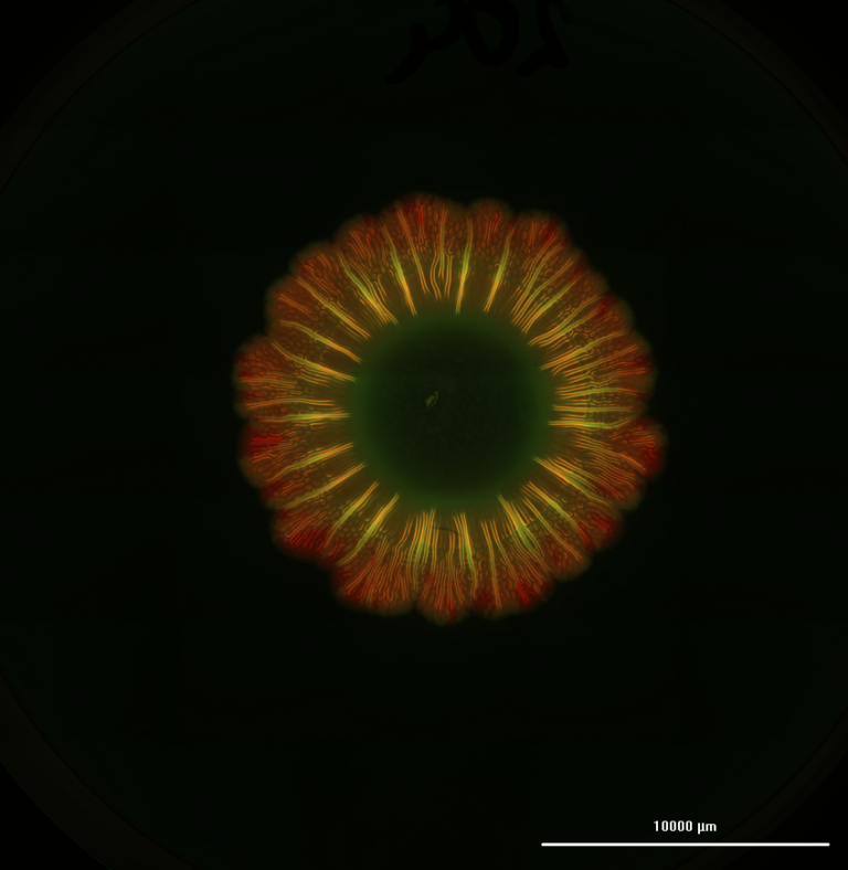
Yes, and an interesting part was finding the right bioink. We used CELLINK’s RGD bioink because it’s functionalized with the short peptides that make cells feel more at home, communicate with each other, and cue each other, but also because it’s relatively clear. When we culture cells in a bioink, we need to monitor their progress. We take bioprints from different time points and examine them under standard fluorescent microscopes. If we cannot see the cells, we have to stop the experiment.

Colin McGuckin
We also don’t want a bioink which has too much complexity for the cancer cells. Sometimes we add adhesion molecules, cytokines or drugs, but we don’t want anything which is going to chemically change the bioink or could cause a reaction towards the cancer cells. Choosing an established bioink supplier like CELLINK ensures we can buy the same bioink again to reproduce results, and there is consistency from batch to batch.
Victoria Sarafian

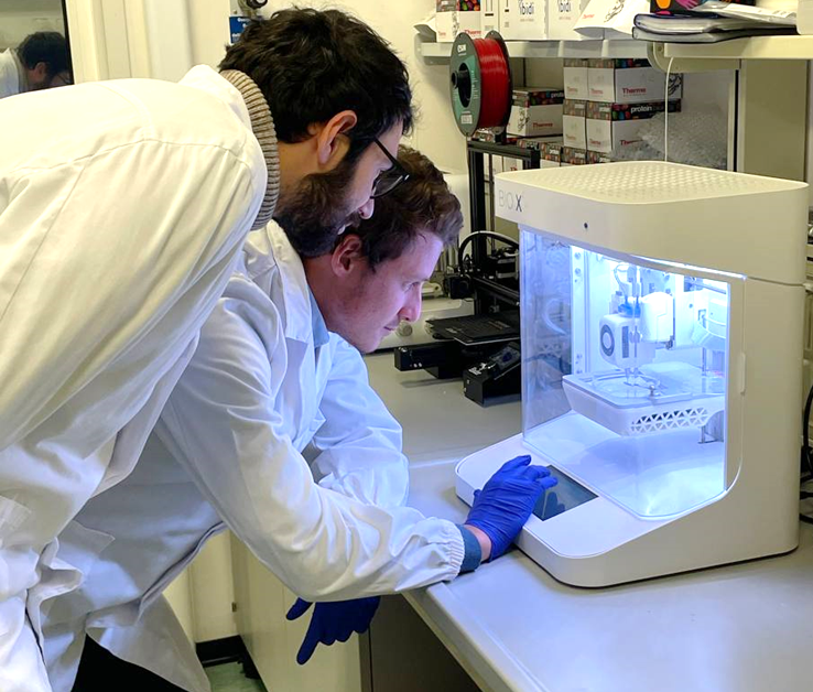
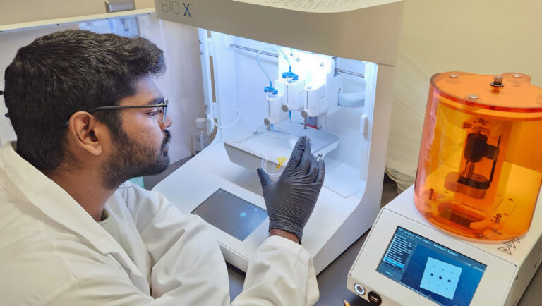
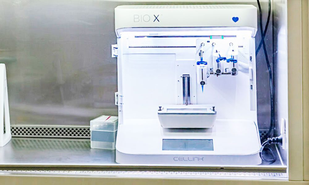
We also used CELLINK’s BIO X™ to bioprint the 3D colorectal cancer models, which didn’t take very long. Then we used several techniques to assess the bioprinted tumors. Tissue staining was used to determine whether these were really the cancer cells we were looking for. Cell viability was assessed at different time points to ensure that our protocol was working well and that we would have the amount of cells needed for the next step. It was a long, tricky process, but we’re quite satisfied with the results we got with the protocol developed with Yordan.

What were some of the challenges unique to colorectal cancer research?

Yordan Sbirkov
Bacterial infection was the biggest bottleneck for growing primary colorectal cancer cells. We were using cells from the patient’s gut, where there are millions of bacteria, some of which become resistant to antibiotics over the years. In the lab, those bacteria grow regardless of what antibiotics you use, and in a few days, cells die.
Colin McGuckin
Our success rate is getting better, however. In Lyon, we are finding ways to expand cells and sterilize them as quickly as possible. Another ongoing project is developing growth media without animal products. We find that the fewer animal products we use and the more defined our media is, the more cells we can grow, and the longer we can grow them. For the first time, we were able to take printed cancer cells and keep them alive for more than 8 months outside the body, and they are still growing, still reproducing. Observing these cells can help us better understand what happens during cancer metastasis.

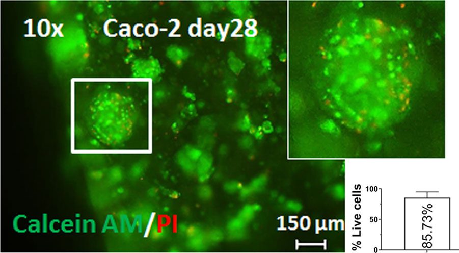
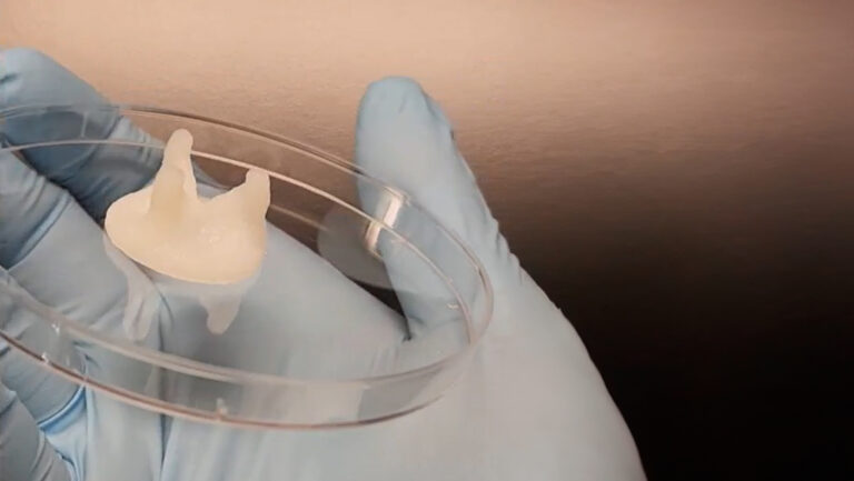
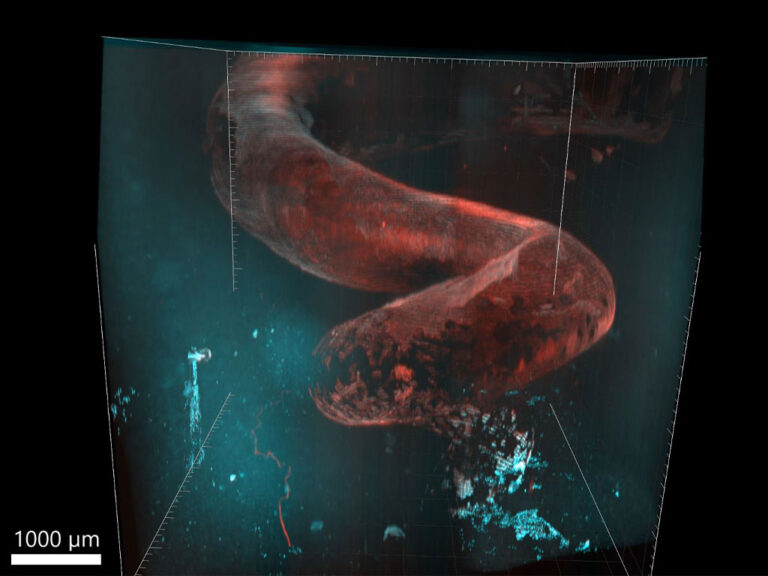
Green staining of live colorectal cancer cells reveals an 85.73% survival rate 28 days after bioprinting on the BIO X.

Which CELLINK service stood out?

Victoria Sarafian
We were very pleased with the friendly, user-focused CELLINK team who installed the BIO X and did the initial training.
Yordan Sbirkov
Before settling on the BIO X, though, we looked at three different bioprinters. Some had overly complicated interfaces with very hard-to-use software, while others weren’t built as well as the BIO X. We found that the BIO X had the best combination of ease of use, the most flexibility to do different things with the bioinks and a better price as well.
Colin McGuckin
I’ve been making 3D tissues for more than 25 years. As an early adopter of new technologies, I was quite pleased with what the BIO X was able to do. A lot of the other options on the market weren’t really bioprinters. Their developers weren’t thinking too much about how living cells would go inside a 3D printer designed to extrude plastics. CELLINK’s approach with the BIO X really focused on the cells and on how to put them into your model. I also appreciated that the software team put out several useful updates along the way. While we don’t need help very often, to be honest, because nothing really goes wrong with the BIO X, CELLINK’s people are always willing to help us with troubleshooting or handling the bioinks. Our experience has been very good, and we now have several BIO X bioprinters in our lab’s bioprinting suite.

Related products

BIO X

This award-winning design is a go-to bioprinter for academics, researchers and innovators.

CELLINK RGD

This bioink provides excellent printability properties and a natural cellular environment with added RGD biofunctionalization.

More Customer Spotlights

Innovative Silk Bioink for 3D Bioprinted Bone Marrow Tissue Models

There is a rising need of platelet units, which is primarily sourced from healthy donors. However, the natural production of platelets coupled with the brief shelf life does not meet the demand by the healthcare industry. Prof. Alessandra Balduini and her team at the University of Pavia are taking this challenge head-on by initiating the EIC Transition SILKink project, which aims to produce a groundbreaking platform that combines the use of natural silk and 3D bioprinting to recreate the environment of the human bone marrow where platelets are produced.

Bioprinting pediatric heart valves that stand the test of time

Heart valve disease is a significant global health issue, affecting millions of people and contributing to a substantial number of cardiovascular-related deaths. Traditional mechanical replacements, while life-saving, present several challenges, especially over time. Most mechanical valves do not grow or adapt with the patient’s body, leading to the need for multiple invasive surgeries. This not only poses inherent risks but also significantly impacts the patient’s quality of life. Issues such as blood clots, calcification, and severe inflammation further complicate the situation. These challenges become even more critical in pediatric cases, where conventional solutions, while still life-saving, may reduce life expectancy by up to 50%. Professor Savoji, Arman Jafari, a PhD student in his lab, and a dedicated team at the University of Montreal have employed bioprinting and their BIO X6 to address these challenges and develop a method to bioprint heart valve replacements.

Empowering Highly Motivated Researchers

We have interviewed Takaaki Arahira, the Associate Professor of the Department of Information Networks, Faculty of Management and Information Sciences at Kyushu Institute of Information Sciences.

Smart Biomaterials: Revolutionizing Drug Delivery

With so many disadvantages having the potential to severely undermine the successful outcome of the therapy, there is an immense requirement, driven by a fusion of both need and convenience, for biomaterials that operate autonomously and intelligently, effectively eliminating the need for human micromanagement. Professor Jeff Bates and his team at the University of Utah are taking this challenge head on. We spoke to Ashwin Velraj, PhD student in Professor Bates’ lab.