3D Bioprinting of Cardiac Tissue: A Protocol for Human-Scale Cardiac Patches
Repairing damaged heart tissue is a complex challenge. While researchers have made significant progress in developing cardiac cells and 3D models, translating those cells into larger, organized tissue constructs remains difficult.
3D bioprinting offers a way to engineer tissue with controlled architecture and composition. But as these approaches move toward larger and more complex constructs, reproducible fabrication becomes just as important as the underlying biology.
A new protocol from researchers at the University of Technology Sydney and the Heart Research Institute provides a detailed workflow for extrusion 3D bioprinting of human-scale cardiac patches containing human cardiac spheroids. Published in STAR Protocols by Niina Matthews and Associate Professor Carmine Gentile, the protocol brings the complete workflow together, from bioink preparation through printing and post-processing.
We spoke with Gentile to learn more about the motivations for and challenges with creating this protocol. “This article brings forward a long-lasting need in cardiac tissue engineering, bringing together over 13 years of research in biomaterials, stem cells and bioprinting technologies, altogether in a protocol to create patient-specific cardiac patches,” he starts.
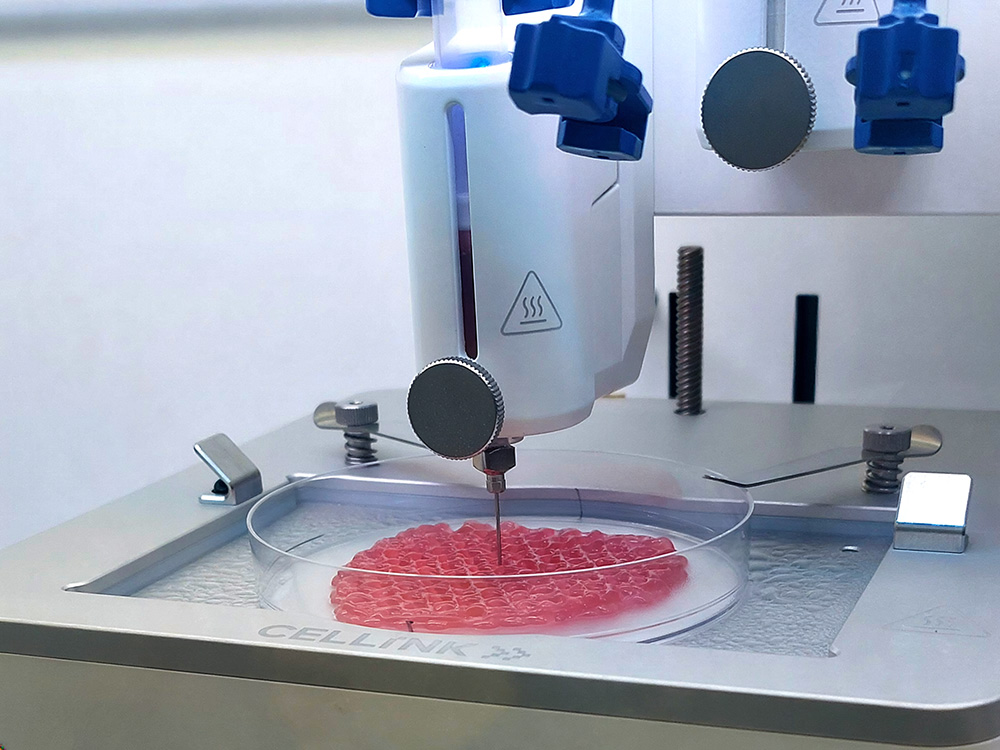
Cardiac patch printed with the BIO X at the Gentile Lab.
Image Copyright: Niina Matthews
From cardiac spheroids to printable cardiac patch
The protocol builds on the Gentile Lab’s work in cardiac tissue engineering and 3D bioprinting, using human cardiac spheroids embedded in a temperature-controlled hydrogel bioink.
The formulation combines 1% silk fibroin, 4% alginate, and 8% gelatin, providing a matrix that can be extruded while supporting the incorporation of cardiac spheroids. The protocol then guides researchers through the parameters needed to produce consistent prints and explains how to troubleshoot common challenges during the extrusion process.
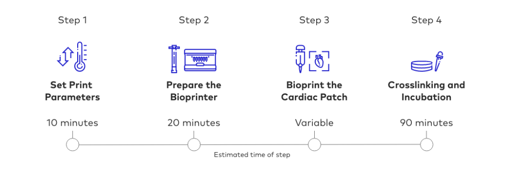
The protocol is designed around a temperature-controlled bioink and provides guidance for optimizing the printing process. Although developed for a specific experimental setup, the authors note that the principles can also be applied to other temperature-controlled biomaterials and extrusion-based bioprinting systems.
“Given the fact that the design of each patch is different from others, as based on the patient’s anatomy, creating a robust and reproducible workflow was quite a challenge. Working with soft hydrogels that are also temperature sensitive made this even more complicated,” Gentile says.
Printing at a relevant scale
As bioprinted constructs become larger, the printing process becomes increasingly complex. Cell viability, print resolution, printing time, and the performance of the final construct all need to be considered together.
Longer printing times can place greater demands on cell viability, while optimizing for high resolution may affect printing speed. At the same time, the behavior of the bioink does not necessarily stop at the printer. Depending on the hydrogel formulation, extruded filaments can swell during crosslinking and subsequent incubation, changing the dimensions and appearance of the final construct.
These considerations become particularly important when moving from small proof-of-concept prints toward larger, more application-relevant tissue constructs. For the Gentile Lab, these challenges were particularly relevant when developing a workflow for human-scale cardiac patches.
“Working with living cells outside of the incubator was a major challenge, while still maintaining hydrogels at the right temperatures during and after bioprinting them,” Gentile said.
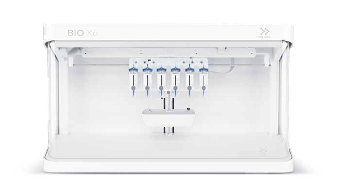
The research team used the CELLINK BIO X6, providing the controlled printing environment needed to work with their temperature-sensitive bioink and optimize the process for larger cardiac constructs.
Building reproducible workflows for cardiac tissue engineering
For bioprinting to contribute meaningfully to cardiac tissue engineering, promising biological concepts need to be translated into workflows that researchers can reproduce and build upon.
“This protocol aims to serve as a way to create personalized patches using the patient’s own scans,” Gentile says before adding, “At the same time, it will help identifying novel methods to explore new hydrogel formulations, as well as explore novel bioprinting methodologies.”
By documenting the process for extrusion 3D bioprinting of cardiac spheroid-laden bioinks into human-scale cardiac patches, Matthews and Gentile provide one such starting point. The protocol combines a defined bioink formulation with temperature-controlled extrusion and practical guidance for optimizing the printing process.
“The ultimate goal is that these patches could be used as heart transplant alternatives, with the hope that they could quickly be translated to the clinic, and save billions of lives globally.”
Further reading
This protocol is part of a broader research program from Dr. Gentile and his collaborators exploring cardiac tissue engineering, 3D bioprinting, and regenerative medicine.
Read more about the lab’s work in our customer spotlight: Mending Hearts on Demand: Bioprinted Patient-specific Heart Tissue
Read the full protocol here: Protocol for extrusion 3D bioprinting of human-scale cardiac patches using cardiac spheroid-laden bioinks