What is 3D bioprinting?
3D bioprinting is additive manufacturing (3D printing) technology that uses biomaterials or biocompatible materials to create 3D tissues and other constructs. The term is an umbrella used to describe many different bioprinting technologies capable of creating biological 3D models.
Within 3D bioprinting, the method of additive manufacturing is an important component, allowing for the positioning of different biomaterials and cells in specific three-dimensional orientations. There are a couple of bioprinting methods that can be utilized, all designed to operate specific biomaterials (usually called bioinks) and different 3D models. We distinguish between two major types of technologies: extrusion-based 3D bioprinting and light-based 3D bioprinting. These technologies are used for many applications within the life science and biotech fields, such as cell-cultured food, drug discovery, personalized medicine, and regenerative medicine.
So, what exactly is 3D bioprinting?
3D bioprinting is a constantly evolving 3D printing technique which uses biomaterials (bioinks) to create 3D tissues or 3D cell culture environments. How a researcher decides to bioprint a tissue model depends on their goals and what they seek to achieve, 3D bioprinting allows for a lot of flexibility to tune the final model to a specific aim. The bioink might be selected to mimic the composition of the tissue that the researcher is looking to print, but it can also be a synthetic formulation developed to emulate a similar function. As mentioned, they may also be printing an environment for culturing cells in 3D and would then choose a bioink or hydrogel suited for that purpose.
To learn more about how bioprinting works, continue reading this article for an in-depth overview of the three main stages: pre-printing, printing, and post-printing.
Bioprinting can be used in an innumerable number of applications, ranging from drug discovery to personalized medicine. Below we dive deeper into these applications and more to give you a better understanding of what 3D bioprinting is.
Learn more about 3D bioprinting:
Bioprinters
Application Notes
Webinars
Our customers’ and partners’ work:
Our customer spotlights
Our customer’s publications
Contact us:
How does 3D bioprinting work?
Describing one exact workflow for 3D bioprinting is tough, as 3D bioprinters, just like 3D printers, provide the user with a lot of freedom to use it for what they are looking to create. If not even more freedom as bioprinters are designed to operate with a lot of different biomaterials. Additionally, 3D bioprinting is a recent invention, and improvements and new technologies are constantly being developed. The application and chosen tissue also heavily affect how you tackle the process. But we can generalize. 3D bioprinting works in three distinct stages: pre-bioprinting, bioprinting, and post-bioprinting.
01.
Pre-bioprinting
At this stage, you own a bioprinter and you have an idea of what you want to achieve in the print. Here you should know what type of model you would like to bioprint, which bioink to use and if you would like to embed cells or not in the 3D bioprinting stage. What you do in this stage is what defines the result at the end of your print session.
If you’re printing with cells (I.E., creating living tissue) you first need to prepare the cells. Once you have the necessary cells, these are mixed* into the bioink you’ve chosen. It may be that you’re developing your own bioink for a specific application. In that case, you will have done the necessary tests on the bioink beforehand to ensure it’s viable for 3D bioprinting.
This is also the time when you develop or select a 3D model. Some 3D bioprinting applications don’t require complicated models – these models can often quickly be generated directly in the bioprinter’s software. Other applications require more complex structures. For example, Ronawk works on developing connecting shapes for iPSC propagation which eliminate the need for subcultures and speed up the cell culture process. These types of complex models are often done in external 3D modeling software but may also be created directly in the bioprinting interface, in a software like DNA Studio 4 for example. No matter how complex the model, they all do the same thing: create a blueprint for the printer to follow in the next stage.
* While the cells are commonly mixed with the bioink, there are also workflows where cells are added at a later stage.
02.
Bioprinting
You’ve created the model and mixed the cells with the bioink – now what? It depends entirely on if it’s an extrusion-based or light-based system.
Bioprinting process: Extrusion-based system
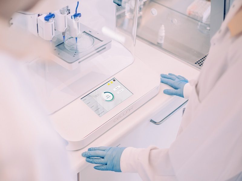
Load your cell-laden bioink into a cartridge and place it into the printhead. On our BIO X series of 3D bioprinters, you can choose between various printheads which have features suitable to use for different types of materials. For example, the Temperature Controlled Printhead is designed to precisely maintain specific temperatures, which is needed when working with biomaterials such as collagen or GelMA. If you’re printing with multiple materials, you may also print with different printheads. This adds another layer of complexity to the bioprinted construct which can lead to a more biomimetic and biorelevant result.
If it’s your first time printing this model with this bioink, you will need to adjust the print parameters. The specific print parameters you should select vary depending on the bioprinter and the bioink properties, for bioinks there is usually a bioprinting protocol provided which outlines which parameters to start at. Once everything is set up and you’ve placed the culture-ware you want to print onto on the print platform, it’s time to press go.
Depending on 3D bioprinter, you can adjust the print parameters as the print is ongoing. This is especially important when bioprinting a specific structure or bioink for the first time, as this allows you to optimize the result without having to restart the print or lose material. Note that, in this phase, it’s often better to not use cells as you don’t want to “waste” them on an unoptimized print.
When the printheads reach the end of your print and stop moving, your print is completed. It now needs to be crosslinked to maintain stable and to lock the bioprinted shape in position. This can be done in multiple ways, the most common approaches are to use photocuring or ionic crosslinking. Thermal gelation and enzymatic reactions are also common to use. Which crosslinking method to select depends on the bioink and the properties of that biomaterial formulation.
Bioprinting process: Light-based system
Every light-based 3D bioprinting system works slightly different. Our layer-less digital light projection (DLP) bioprinter, BIONOVA X works differently from our other series of DLP bioprinters, LUMEN X. What they have in common however is that they utilize light projection to shape the 3D models, allowing for precise reconstruction of structures.
With the BIONOVA X, you print the models directly in well plates. This means that you must load the system with a printing probe that matches the number of well plates. Instead of loading the bioink into a cartridge, you add the bioink into the wells of a well plate. You then proceed to use the bioprinter’s software to load the printer with the well plate and upload the model that is intended to be printed.
The LUMEN X series of bioprinters works like a traditional layer-by-layer DLP printer. Instead of the printing probes used in the BIONOVA X, you attach a build plate to the system. Here, you also don’t use a cartridge. Instead, you place the bioink in a vat, which you then put into position on the printer.
After you’ve set up the bioprinter, it’s time to start. Like how it works for extrusion-based bioprinters, you need to set up and optimize the print parameters. Again, the parameters you choose depend on the printer, the construct, and the materials you choose. Once the model is loaded into the system and parameters are selected, it is time to hit “Print”.
In light-based bioprinting the selected printing parameters are locked in for the duration of the print. To optimize a print, print parameters are changed after a construct is printed and evaluated. If you’re noticing that something isn’t working the way you want, you may also interrupt the process as it is printing and adjust accordingly.
Combining the different 3D bioprinting processes
Some applications can benefit from both light- and extrusion-based 3D bioprinting. Which method to use depend on the material intended to be used as well as desired model characteristics. In general, light-based technologies provides higher resolution and can create finer shapes, while extrusion-based bioprinting is compatible with a wider library of biomaterials and better suited for multi-material bioprinting. The two technologies can be combined, to utilize the strengths of both technologies by moving constructs from one printing technology into the other system.
03.
Post-bioprinting
Your bioink and cells (if included) has been printed into a construct, the construct has been cured and is ready to be used – what you do next depends entirely on your goals. First, you determine that the quality of the print is what you expected. Typically, you then proceed with surrounding the printed construct with cell medium and subsequently place it in an incubator. If cells are embedded in the bioprinted model it is recommended to provide cell media as soon as possible after the print to start the culture phase.
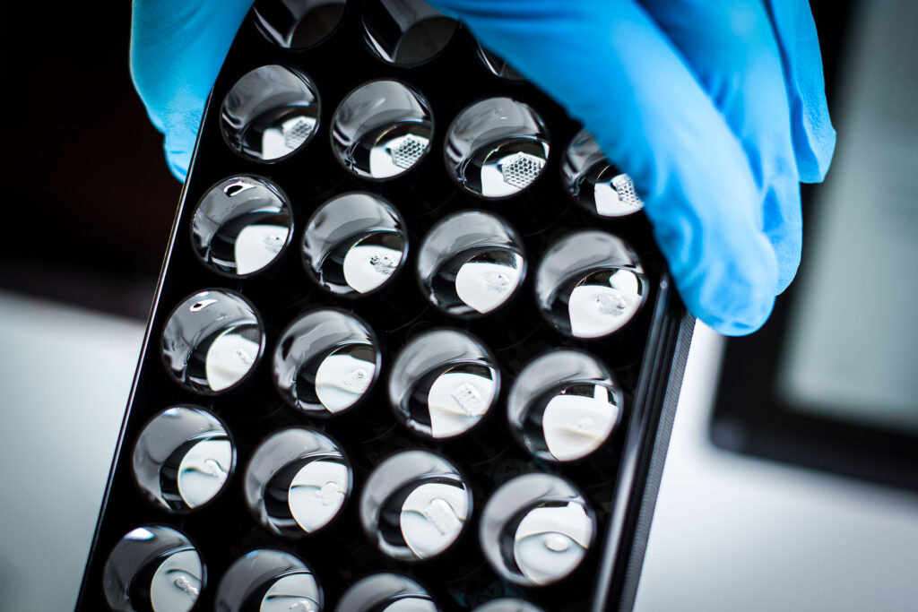
The cell media is like the blood in the body, by suppling fresh media, and including different factors, the 3D bioprinted cell model is matured into the right type of tissue. Once the cell culturing is complete, it’s time to use the construct for what you intended. You may have printed skin and now want to test how your cosmetic product affects it, as a great alternative to animal testing. You may have printed a 3D model into well plates to test various drug candidates efficacy, to faster pick out promising options.
How bioprinting works, in short
3D bioprinting works in many ways as it changes depending on the application that you’re planning on using it for, and the technology you’re using. There are three basic stages of 3D bioprinting that most workflows follow: First, select and prepare a bioink and create a model fit for your application. Second, put the bioink into its correct place for the bioprinter, select the printing parameters, print the construct. Third, treat the 3D construct according to your application.
Bioprinting applications - what are they?
An exact list of applications is impossible to provide, and we’re happy that’s the case. Bioprinting, biotech, biology… They are all fields which are constantly growing, and where science is developing. At CELLINK we’re not making it a secret: our developers and researchers are hard at work ensuring that, in the not-too-distant future, our customers may one day print fully working internal organs. But that’s not all that 3D bioprinting is. From helping regrow the coral reefs and heavily reducing the need for animal testing, to allowing pharma companies to determine promising drug candidates faster through biomimetic cell culture. Below is a list of some 3D bioprinting applications which have shaped what bioprinting is today, and which will shape what bioprinting is in the future.
Drug discovery and development
Developing drugs is an expensive, long, and arduous process. There’s a lot of safety, as well as trial and error, which go into making a new drug. Today, 3D bioprinting doesn’t have the ability to fully eliminate the use of human trials. However, bioprinted constructs are suitable for pharma companies to use in the initial stages of drug development where they need to scale down leads to a few potential candidates due to their biomimetic properties. Access to more biorelevant models at the in vitro testing stage allow the researchers to determine a drug candidate’s efficacy sooner, speeding up the process of finding which compounds to bring to the next stage. As a result of speeding up the process, 3D bioprinting also reduces the cost of drug development. Read more about how bioprinting is being used in drug discovery here.
3D cell culture
3D cell culture is an excellent alternative to traditional cell culture methods. Traditional cell culture is done on a two-dimensional plane, which means that cells are interacting with each other in two dimensions. By adding a third dimension, usually by combining the cells in a biomaterial serving as the ECM of the cells, we allow cells to interact with each other in the manner they would in real life in vivo. What this means, overall, is that 3D cell culture methods allow researchers to study cellular systems in a more native setting and access data that’s more relevant to the conditions of the real world.
3D bioprinting allows you to guide the process of making 3D cell cultures in a way that other 3D cell culture methods have a tough time doing. This means you can use more assays and protocols that are fully tailored to the research you are doing.
Personalized and regenerative medicine
Regenerative medicine is about amplifying and aiding the natural healing process, or about replacing the function of damaged tissue. Personalized medicine is about moving away from the one-size-fits-most healthcare and toward targeted treatments tailored to the individual patient. As our understanding for biological processes advances it has also been identified how important individual profiles, such as genomic or metabolomic fingerprints, are for the efficiency of treatments.
3D bioprinting enables both for very similar reasons. An example of bioprinting for personalized and regenerative medicine are corneas. Multiple companies and research groups are working on 3D printing corneas using specialized inks and stem cells. 3D bioprinting has allowed these companies to print corneas in the specific form they need, ready to be implanted. Effectively, this reduces the reliance on cornea donations around the world. While not fully in use yet – we soon expect this to be used in real life.
Another example is within how biomaterials can help aiding regrowth, both in regenerative medicine as well as in the environment. We are looking at the regenerative properties of biomaterials and bioprinted constructs to help regrow coral reefs, and at University of Alberta researchers look at bioprinted-to-fit nasal cartilage grafts for use in cancer patients who’ve developed nasal lesions.
Read more in the areas of impact section for specific examples on how 3D bioprinting is being used for personalized and regenerative medicine today.
And many more
As mentioned at the start. It is impossible to pin down an exact list of applications, so we won’t even try. If you’re interested in 3D bioprinting technology and are wondering if it is for you, don’t hesitate to contact us!


