Lattice Scaffolds in Bioprinting for Tissue Engineering
Lattice scaffolds are crucial in tissue engineering, providing structural support that closely mimics natural extracellular matrices (ECM). With modern DLP 3D bioprinting technologies, it’s possible to fabricate highly porous, interconnected lattice structures tailored to the mechanical and biological requirements of specific tissues. Advanced designs such as triply periodic minimal surfaces (TPMS) and polyhedral structures like buckyballs offer significant advantages, including improved cell proliferation, vascularization, and integration with host tissues.
Triply Periodic Minimal Surface (TPMS)
Gyroid and Related designs
Triply periodic minimal surfaces (TPMS), particularly gyroid structures, feature continuous, smooth surfaces that partition space into interwoven pore networks, optimizing mechanical performance and cellular behavior. These surfaces exhibit high stiffness and strength compared to traditional lattices, largely due to their smoothly curved architecture that evenly distributes mechanical stress. The BIONOVA X, with its in-motion printing mode, ensures smooth and continuous biomaterial photocuring, printing the intricate curves and seamless surfaces characteristics of gyroid scaffold.
Gyroid structures particularly demonstrate isotropic mechanical properties, making them ideal for load-bearing applications such as bone regeneration. The gyroid’s open and interconnected pore network greatly enhances cell proliferation and viability. Studies have shown superior cell seeding efficiency and homogeneity in gyroid scaffolds compared to random porous structures. The geometry itself can stimulate cell behavior via mechano-transduction pathways, enhancing osteogenic differentiation and promoting robust vascularization. This interconnected porosity ensures nutrients and oxygen efficiently reach cells throughout the scaffold, significantly improving tissue regeneration.
Gyroid scaffolds have shown notable success in promoting vascular infiltration, critical for tissue engineering. In vivo studies have observed rapid vessel ingrowth, highlighting the scaffold’s potential in supporting large tissue constructs that require efficient nutrient delivery.
Additionally, gyroid scaffolds demonstrate excellent tissue integration, particularly in bone regeneration, supporting firm anchoring with minimal fibrous encapsulation. This integration is further enhanced by controlled scaffold degradation, ensuring gradual and uniform replacement by native tissue.
Spherical Polyhedral Scaffolds
Another notable design inspired by nature is the spherical polyhedral lattice, specifically buckyball-like scaffolds. These structures resemble truncated icosahedra, characterized by hexagonal and pentagonal openings.
Buckyball lattices are primarily used as micro-scaffolds for encapsulating cell spheroids, facilitating modular tissue engineering. Although individually these structures are modestly mechanically robust, their spherical geometry provides significant geometric rigidity, beneficial for preserving structural integrity during tissue culture and handling.
Buckyball scaffolds excel as microenvironmental regulators, promoting robust cell growth and maintaining the differentiation potential of encapsulated spheroids. The cages allow controlled cell aggregation, preventing over-compaction or uncontrolled merging typical in scaffold-free approaches. Cells cultured within these micro-scaffolds exhibit enhanced nutrient diffusion and better maintenance of spheroid shape, ensuring uniform tissue assembly in a modular manner.
While direct studies on vascularization within buckyball scaffolds remain limited, their inherent openness and large apertures theoretically support excellent perfusion and capillary penetration. The modular nature of buckyball structures further aids integration with host tissues by allowing smaller tissue units to merge seamlessly, enhancing overall tissue continuity. Early in vivo studies show encouraging results in cell survival and initial matrix deposition, suggesting significant potential in tissue regeneration applications.
Other Novel Lattice Designs
Thanks to their mathematical versatility, lattice structures offer virtually unlimited design possibilities enabling precise control over mechanical, transport, and biological properties in printed constructs.
Schwarz-P Scaffolds
Schwarz-P triply periodic minimal surface (TPMS) scaffolds are increasingly used in bone tissue engineering due to their highly interconnected, smooth pore architecture that enhances fluid permeability, cell migration, and mechanical stability. Their mathematically defined geometry allows precise tuning of porosity and pore size to match the needs of cortical or cancellous bone, with permeability often outperforming other TPMS designs.
Beyond facilitating nutrient flow and osteointegration, Schwarz-P lattices demonstrate exceptional structural integrity, even after microscale fabrication distortions, exhibiting mechanical moduli closely aligned with bone and variances of less than 7% from experimental value.
Voronoi Stochastic Scaffolds
Voronoi-based stochastic scaffolds, which emulate natural trabecular bone, feature randomized pore sizes and orientations, closely matching the irregular structure of natural bone. These biomimetic structures demonstrate enhanced osseointegration and can replicate the anisotropic mechanical properties characteristic of natural bone, offering customized solutions for patient-specific needs.
As the BIONOVA X bioprinter is optimized for cellularized scaffold printing, it is essential to bring the condition for cells growth after printing by providing optimal pore distribution. Recent studies emphasize the critical importance of pore size and geometry in tissue outcomes, indicating there is no universal optimal pore dimension.
Smaller pores (~100–300 µm) are beneficial for initial cell aggregation and bridging, while larger pores (>300 µm) facilitate vascularization and deeper tissue infiltration. Advanced lattice scaffolds combine these pore scales, optimizing early cell seeding and subsequent vessel ingrowth.
Additionally, biofunctionalization of lattice surfaces with bioactive molecules or extracellular matrix components is enhancing cell adhesion and guiding tissue-specific responses.
Gradient and Multi-scale Lattices
Gradient and multiscale lattices represent another innovative approach. These scaffolds gradually vary pore sizes or combine macro and microscale pores, facilitating seamless integration of different tissue types, such as cartilage transitioning to bone.
The controlled gradients direct specific cell types to suitable environments within the scaffold, enhancing tissue-specific regeneration while maintaining structural integrity. Moreover, the flexibility of BIONOVA X allow a fine tuning of parameters to adjust sections based on the level of complexity of the structure bring the properties required in term of matrices stiffness thanks to grayscale bioprinting option.
Recent advancements in computational design and artificial intelligence are driving innovation in lattice scaffold engineering. Topology optimization and machine learning algorithms now enable the creation of optimized lattice structures tailored to precise mechanical and biological criteria, significantly improving tissue regeneration outcomes.
In vivo studies of advanced lattice designs are encouraging, demonstrating successful load-bearing capacities and effective bone regeneration with minimal inflammation. Pre-vascularized modules using buckyball-inspired scaffolds have shown potential in forming vascularized tissue constructs. Such developments suggest lattice scaffolds will play a vital role in future tissue-engineered implants.
Multi-material Lattice Scaffolds
Emerging applications for lattice scaffolds now extend beyond bone and cartilage regeneration to cardiac and neural tissue engineering. Specifically engineered lattice structures can direct muscle or nerve cell alignment and enhance functional integration, highlighting the versatility and potential of these designs in regenerative medicine.
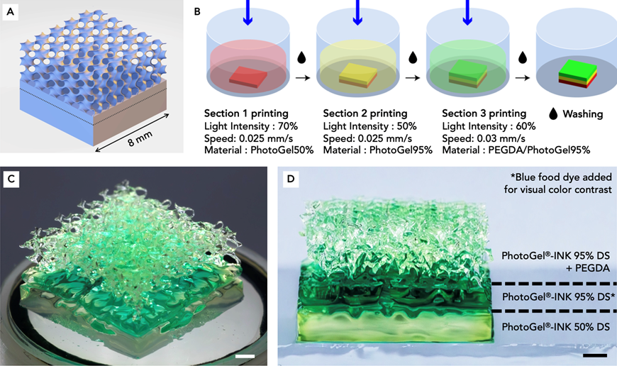
In this context, multi-material bioprinting techniques are expanding possibilities, allowing combinations of stiff supportive materials and bioactive hydrogels within the same scaffold structure. Such hybrids provide immediate mechanical stability alongside bioactivity crucial for cell proliferation and differentiation. Bioprinting with multiple hydrogels allows researchers to more accurately recapitulate the complexity of native human tissue, where different regions may vary in mechanical properties, cell composition, and ECM structure.
Multi-material bioprinting offers several advantages for replicating complex anatomical and functional features of living human tissue:
- Modeling of heterogenous tissue, such as osteochondral interfaces and skin.
- Spatial cell placement, with different cell types embedded in distinct hydrogel regions.
- Controlled biochemical environments, by localizing growth factors, peptides or drugs within specific zones.
- Improving physiological relevance for regenerative medicine and in vitro drug testing.
In addition to multi-material printing, the BIONOVA X also allows for tunable stiffness gradients, enabling mechanical transitions that match native tissue, further recapitulating the native tissue environment.
Conclusion
In summary, lattice scaffolds have fundamentally transformed tissue engineering by offering precise control over both the mechanical environment and cellular responses. Advanced structures like TPMS gyroids and buckyball cages actively promote cell viability, differentiation, and tissue integration, making them promising candidates for regenerating complex, vascularized, load-bearing tissues. As research continues to advance, we anticipate the development of increasingly sophisticated and dynamic lattice designs tailored to individual clinical needs, paving the way for effective and functional tissue regeneration.
References
Ataollahi, S. et al. (2023). Additive manufacturing of lattice structures in tissue engineering: Materials, designs, and strategies. Bioprinting, 35, e00304. – A comprehensive review of lattice scaffold fabrication techniques and designs, highlighting the roles of different unit cell geometries in tissue regeneration.
Yuan, X. et al. (2023). The design of strut/TPMS-based pore geometries in bioceramic scaffolds guiding osteogenesis and angiogenesis in bone regeneration. Mater. Today Bio, 20, 100652. – Demonstrated that gyroid pore scaffolds enhanced HUVEC endothelial network formation (CD31 expression, tube formation) and activated mechanotransductive pathways (YAP/TAZ) to promote vascularized bone regeneration.
Diez-Escudero, A. et al. (2020). Biomimetic scaffolds using triply periodic minimal surface-based unit cells: design, fabrication, and evaluation. Biomaterials, 232, 119719. – Evaluated various TPMS (gyroid, diamond, etc.) in polymer scaffolds, reporting superior mechanical properties and cell attachment for TPMS vs traditional lattices.
Li, J. et al. (2023). 3D-printed gyroid scaffolds with bioactive fillers for bone repair: in vivo osteogenic and angiogenic assessments. Biofabrication, 15(4), 045012. – Showed that a DLP-printed gyroid scaffold with isosorbide-based polymer and hydroxyapatite had quick vascular ingrowth (vessels infiltrating by day 7) and significant new bone formation in a rat calvarial model.
Kopinski-Grünwald, O. et al. (2024). Scaffolded spheroids with enhanced self-assembly dynamics as building blocks for bottom-up tissue engineering. Acta Biomaterialia, 174, 163-176. – Introduced microscopic buckyball-shaped scaffolds to house stem cell spheroids. Found that microscaffolds preserved cell viability and differentiation potential, while improving cell retention and uniform tissue size during assembly.
Kanwar, S. et al. (2023). Additively manufactured stochastic and gyroid scaffold design towards osseointegration and bone regeneration in a rabbit femur model. Mater. & Design, 235, 111723. – Compared a biomimetic random (stochastic) porous scaffold to a periodic gyroid scaffold in vivo. Both supported bone ingrowth, but the stochastic design better replicated natural bone architecture, highlighting the benefit of Voronoi-like lattice in certain scenarios.
Melchels, F. et al. (2010). Effects of the architecture of tissue engineering scaffolds on cell seeding and culturing. Acta Biomater, 6(11), 4208-4217. – Pioneering work showing mathematically designed scaffolds (including TPMS) achieve more homogeneous cell seeding and distribution than random porous scaffolds, due to controlled pore size and connectivity.


