3D human liver model using GelXA Laminink

IN VITRO STRUCTURAL 3D LIVER MODEL


Hyun Kyoung Kang is a Ph.D student working in Professor Kyung-Sun Kang’s laboratory in the college of Veterinary Medicine at Seoul National University, Republic of Korea. They are investigating direct reprogramming from fully differentiated somatic cells to neural stem cells. By understanding the basic role of stem cells, they are trying to apply new therapeutic approaches to inveterate diseases. Additionally, they research tissue engineering and brain/skin organoids.

Aim

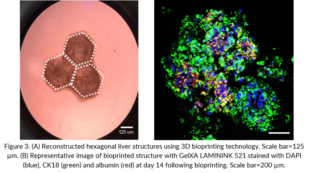
This project was conducted at Seoul National University (SNU) with bioinks provided by CELLINK. The SNU team constructed a model of hepatic tissue that resembles hexagonal liver lobules using 3D bioprinting technology. The aim of this collaboration was to enhance and develop the technology of 3D tissue bioprinting, particularly creating a 3D in vitro human liver model using hepatic cells with different types of GelXA LAMININK bioinks.


The G-code to this three-layered grid structure can be found on Bioverse!

Bio X printing of HepG2
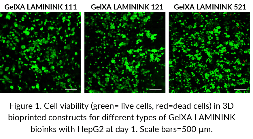
For this liver tissue model, the bioinks CELLINK LAMININK 111, 121, and 521 were used to create specialized environments for the maturation of the cells. The evaluation focused on the compatibility of the bioprinting process and encapsulation of the HepG2 cells. 3 million cells/mL bioink was bioprinted as grid patterns and cultured for a month. The cell viability was tested after one week using Calcein AM/ethidium homodimer reagents. After 14 days following bioprinting, the bioprinted structures were analyzed with hematoxylin and eosin (H&E) staining, and the expression patterns of hepatocyte makers were analyzed with immunostaining.

Viability

A live/dead assay with Calcein AM and ethidium homodimer was performed on bioprinted structures generated in different types of GelXA LAMININK bioinks. Seven days post-bioprinting, the cell viability of HepG2 cells in constructs bioprinted with GelXA LAMININK 111, 121, and 521 was compared (Figure 1). Interestingly, the HepG2 cells displayed the highest cell viability in GelXA LAMININK 521. This indicates that laminin 521 could be a favourable additive for increasing HepG2 viability, and thus GelXA LAMININK 521 is an appropriate bioink for bioprinting in vitro liver models.

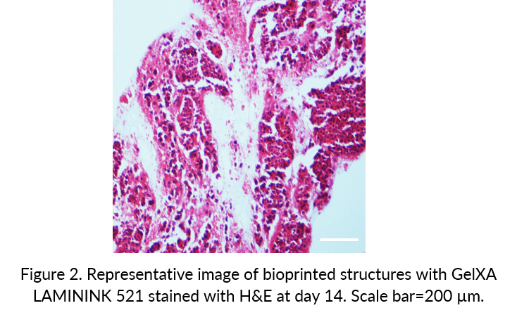
Well distributed cells

After 14 days, the bioprinted constructs were fixed with 4% formalin, embedded in paraffin, cut into 10 μm thick sections, and stained with H&E. H&E staining of the bioprinted constructs verified cell position and morphology (Figure 2). HepG2 cells were initially well-distributed but self-aggregated to form a tissue.

Albumin expression

The liver exhibits a hierarchical structure consisting of repeated functional tissue units (liver lobules). In this regard, the hexagonal hepatic structures were designed to be printed in order to replicate the anatomical structures of three hepatic lobules (Figure 3A). Here, immunostaining confirmed the existence of non-secreted protein, CK18, and representative hepatocyte marker, albumin (Figure 3B). This demonstrates that HepG2 cells express hepatocyte markers while embedded in bioprinted constructs.

Conclusion

Construction of liver structures using a 3D bioprinter could provide an alternative strategy for modelling of the human liver in vitro. HepG2 cells were mixed with three different GelXA LAMININK bioinks and then bioprinted using the BIO X. The SNU team conclude that GelXA LAMININK 521 enables HepG2 cells to grow efficiently in 3D culture conditions. The bioprinted structures presented here highlight the successful application of 3D bioprinting technology to liver tissue engineering.