In this video, we printed droplets with a 22G nozzle. We mixed CELLINK Bioink with cell suspension in a 1:10 ratio. Using 10-15 kPa pressure, we extruded single droplets for 0.75 seconds on a 96-well plate.
Add our Crosslinking Agent to ensure the stability and longevity of your printed constructs. After washing with medium, your cell-laden droplets are ready to be cultured in a standard cultivation environment.
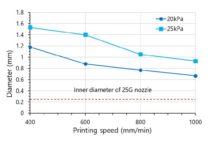
Low pressure + high speed = Great printability
The Droplet function easily prints every bioink we offer, but bioprinting conditions and bioink properties can affect filament extrusion.
CELLINK Bioink produces a continuous filament through a wide range of nozzle diameters. Combine low pressure with high print speed to minimize the difference between the inner nozzle diameter and extruded filament width (Figure 1). CELLINK Bioink contains nanocellulose, and we recommend using a minimum nozzle diameter of 25G to prevent clogging. Change the nozzle size, pressure or extrusion time to control droplet volume.
Figure 1. Filament width dependence on a printing speed and pressure using a 25G nozzle
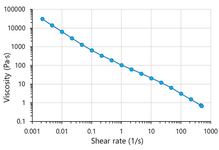
The perfect shear-thinning bioink
CELLINK Bioink contains well-dispersed cellulose nanofibrils that form a stable network. CELLINK Bioink’s viscosity is indirectly related to shear rate, making it an excellent material for bioprinting (Figure 2).
The filament extrudes smoothly and maintains shape after printing. Temperature doesn’t affect CELLINK Bioink’s performance, so you can print with ease at room temperature.
Figure 2. Viscosity dependence on shear rate for CELLINK Bioink
Generating 3D tissue models
Leverage the Droplet function to create droplets with one or more cell types and generate high-throughput 3D tissue models with high reproducibility.
Bioprinted tissue models can mimic the native phenotype of any cell type. They have the potential to aid drug discovery with high-throughput drug screening, improve primary cell survival for cells requiring controlled suspensions and enhance understanding of cellular communication and maturation.