"Studying the ill-defined cross-talk between the leukemic cells and their microenvironment"
Our customer, associate professor Cristina Scielzo at San Raffaele Scientific Institute in Italy, is looking at primary leukemic cells’ cross-talk with their microenvironment. For this to be a good pathological-like in vitro model, she is 3D printing the cells in the CELLINK’s LAMININK bioinks and CELLINK’s RGD bioink. Read her testimonial here below.
“The focus of my research is to study the ill-defined cross-talk between the leukemic cells and their microenvironment. To study in depth, the nature of this cross-talk, we aim to re-construct a more physio pathological-like in vitro model using the 3D bioprinting strategy.”
“We are taking advantage of primary cells isolated from leukemia patients’ or healthy donors’ peripheral blood. We are mixing the cells with bioinks specifically designed to support cellular attachment and functions. The CELLINK’s LAMININK bioinks demonstrates high printability and biocompatibility.
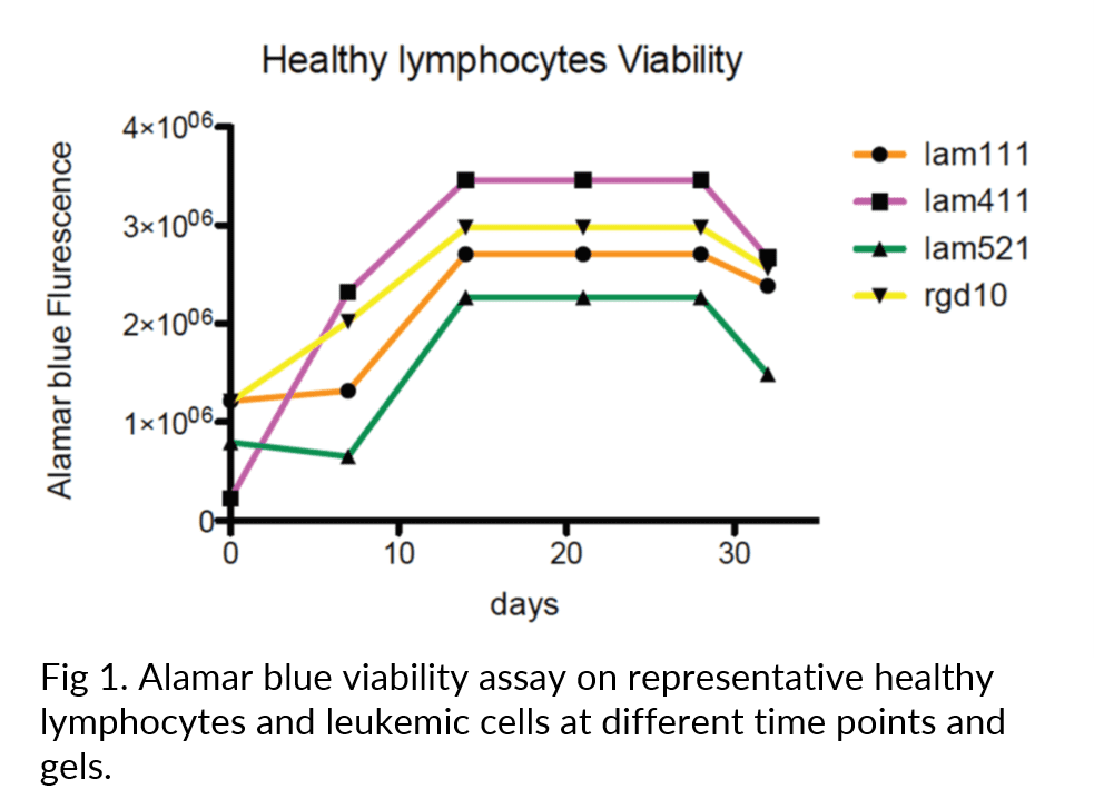
In particular, with the aim to recreate different stimuli derived from the extracellular matrix, we are testing in parallel different bioinks within the CELLINK’s LAMININK bioinks and comparing to CELLINK’s RGD bioink. We successfully tested leukemic cells for printability, behavioral change and viability. We optimized the printing strategy and set-up protocols for performing analysis for cells viability, flow cytometry, RNA extraction, protein extraction and confocal microscopy. We found that leukemic cells have a long-term viability (up to 21 days) in the 3D bioprinted scaffolds (Figure 1).”
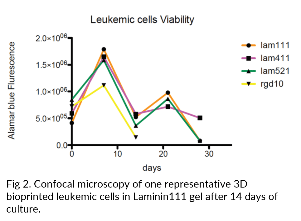
“Finally, using the CELLINK CELLINK’s LAMININK bioinks we found a different localization pattern of the cells, in particular more aggregates were observed with CELLINK’s LAMININK 111 bioink (Figure 2).
Our preliminary results show that we can efficiently 3D bioprint and follow leukemic cells over time in 3D allowing for more reliable physiological cell-cell interactions. Moving from a 2D culture system to more challenging 3D method, we will be able to study the behavior of leukemic cells in relationship to other cells and different extracellular matrix at basal level and when exposed to anti-leukemic compounds.”