Evaluating Liver Toxicity in Bioprinted Mini Livers
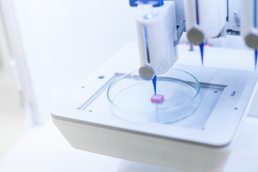
Application note covers studying cytotoxicity in bioprinted liver models. Liver models biofabricated on the BIO X bioprinter demonstrated cytotoxic responses to drugs. Findings further validate 3D bioprinting as a viable high-throughput solution for modeling hepatic tissue and screening for drug-induced liver injury (DILI) and idiosyncratic drug reactions. In vitro 3D liver models can replace costly and low-throughput 2D cell culture systems and animal studies. The new method of “droplet in droplet” (DID) bioprinting uses the BIO X to droplet-print hepatic (HepG2 and LX2) and non-hepatic (HUVEC) cells encapsulated in type I collagen.
Learn how:
- To produce functional 3D bioprinted mini livers as reliable alternatives to 2D cell culturing, animal studies or lab-on-a-chip prototypes.
- Studying cytotoxicity in bioprinted liver models enables high-throughput drug screening and analysis.
- Incorporating type I collagen for DID models provides a highly compatible environment with abundant binding sites for hepatic cellular rearrangement and spheroid formation.
- A DID model allows for cell-cell interactions between layers of tissue and a unique opportunity to investigate migration patterns between layers.


