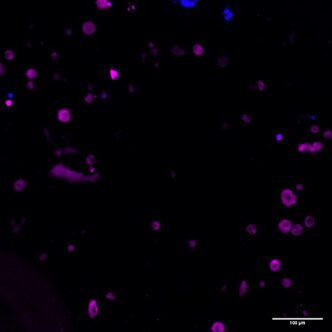
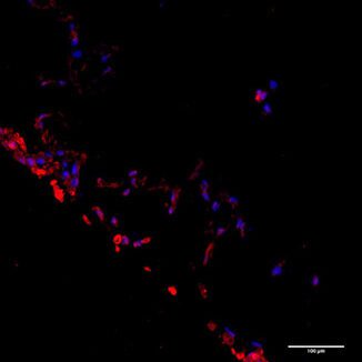
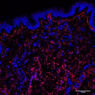
Successfully bioprinting skin tissue models requires overcoming two challenges: 1) producing physiologically functional tissue and 2) comprehensively analyzing that tissue with specific antibodies. With CELLINK’s Skin Tissue Model Kits, surmounting the former is possible with the use of bioinks explicitly developed to support the maturation and proliferation of cells that produce the thick dermis layer and those that create the stratified epidermis. The latter is dealt with by deliberately optimizing antibodies that express cell phenotypes in the created skin models.