Liver tissue model kits
Liver tissue modeling just got a whole lot easier!
CELLINK’s Liver Tissue Model Kits provide all the tools necessary to generate bioprinted liver tissue models and perform a complete analysis. Such liver models maintain a physiological 3D environment that resembles in vivo conditions, which enhances liver cell processes. As a result, liver tissue models demonstrate native-like uptake and dose responses, enabling, for instance, more relevant drug screening. The complex bioinks in the Liver Tissue Model Kits help tailor liver models for specific investigations and the optimized antibodies allow for direct analysis of the model’s cells with selective key markers for liver functionality.
Two bioink options
CELLINK Bioink or GelXA-based bioinks functionalized with laminin 111 are provided in the Kits. The GelXA LAMININK 111 provides the biological properties of GelMA with printability at a wide temperature range, including room temperature. It has crosslinking capabilities to better accommodate cellular sensitivity, allowing you to tune mechanical characteristics of the models. CELLINK LAMININK 111 is advantageous with stable and versatile printability characteristics at any temperature, in addition to not being sensitive to light. The nanocellulose in the CELLINK LAMININK 111 provides a fibrillar network that can influence the interconnections of the different liver cell types, potentially facilitating, for example, the formation of a bile canalicular network or the alignment of stellate cells within a co-culture tissue model.
Liver tissue model kit
Complete solution for generating and analyzing liver tissue models. The kit comes with bioinks that accommodate multiple cell types including hepatocytes, stellate cells, Kupffer cells and endothelial cells, giving you the ability to tailor each model to your needs
Three antibodies
Direct functional analysis of the liver tissue model can be obtained through immunohistochemical imaging of the three antibodies collagen type I, ABCC2 and CYP3A43. The collagen type I antibody evaluates the production of collagen type I by the stellate cells during fibrotic inductions. The ABCC2 antibody evaluates the transporter multidrug resistance protein 2 (MRP2) in hepatocytes, while the CYP3A43 antibody analyzes one of the cytochrome P450 enzymes responsible for metabolism of drug compounds.
Liver tissue model kits in action
Antibody in green, DAPI in blue, scale bar = 100 µm.
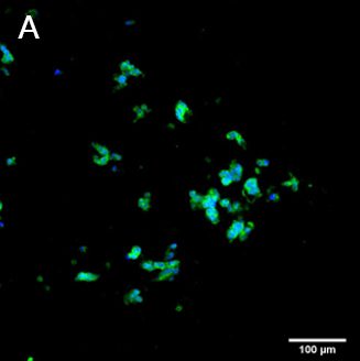
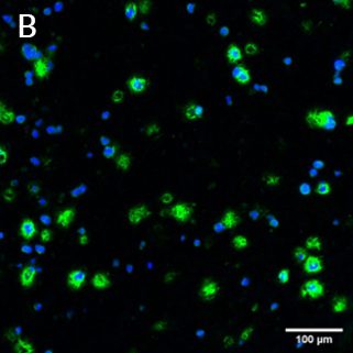
Collagen type I production in co-cultured HepG2 and LX2 in (A) GelXA LAMININK 111 and (B) CELLINK LAMININK 111 at Day 7 of culture.
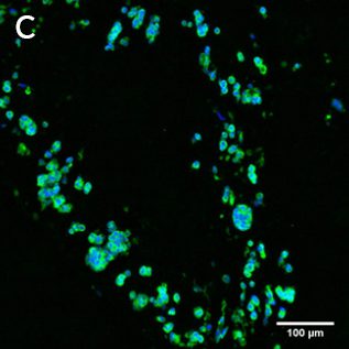
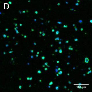
The ABCC2 antibody staining of hepatocyte HepG2 in co-cultured constructs bioprinted with (C) GelXA LAMININK 111 and (D) CELLINK LAMININK 111
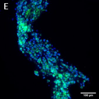
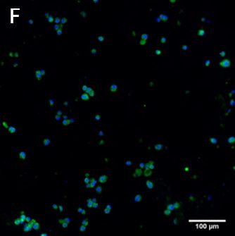
The CYP3A43 antibody staining of primary hepatocytes in (E) clusters and (F) single cells.


