Applications / Spheroids and Organoids
Spheroids and Organoids
Design Reproducible, Assay-ready 3D Models with Bioprinting
Spheroids and organoids are essential 3D cell culture models that reproduce key features of in vivo environments for research, including drug discovery and disease modeling. Integrating bioprinting into the workflow adds control over model placement, architecture, and microenvironment, enabling scalable and more physiologically relevant assays. This controlled approach supports greater consistency and reliability while maintaining compatibility with standard culture, imaging and analysis workflows.
Day 14 brightfield image, showcasing spheroid formation of A549 lung cancer cells in small droplets of GelMA C bioink.
What are oids and why are they important?
Spheroids are compact multicellular aggregates that reproduce key cell-cell interactions and tissue-like gradients. Organoids are self-organizing 3D models that reproduce selected structural and functional features of an organ. They both provide a more biologically relevant context than conventional 2D culture and are used across fundamental research, disease modelling, and drug development. When designed and validated for a defined context of use, human-relevant spheroids and organoids can serve as New Approach Methodologies (NAMs) for studying human biology, disease mechanisms, and treatment responses.
HOW CELLINK CONTRIBUTEs TO
Transforming spheroids and organoids into better research models
CELLINK bioprinters help researchers move from manually prepared 3D cultures to more controlled experimental formats. Cells, preformed spheroids or organoids, and supportive biomaterials can be deposited in defined positions and geometries, improving consistency across wells, plates and experimental runs.
Researchers can standardize deposition volume and model geometry, organize multiple cell types or compartments, and tailor matrix composition, stiffness and biochemical cues to the biological question. Printing directly into commonly used plate formats also supports repeatable microscopy, longitudinal monitoring and treatment-response studies.
The goal is not simply to generate spheroids and organoids, but to create more reproducible, informative and assay-ready research models. CELLINK supports these workflows through biodispensing systems, extrusion and light-based bioprinters, and a portfolio of biomaterials designed for spheroid, organoid, and advanced 3D cell culture applications.
Research Areas
From Controlled Information to Application-ready Models
Cancer biology and drug screening
Generate tumor spheroids or organoids for treatment-response studies, quantitative imaging and advanced tumor-microenvironment research.
Disease and development biology
Create tissue-specific models with 3D bioprinting to study development, disease mechanisms, and cell-matrix interactions.
Patient-relevant and functional testing
Regenerative medicine and tissue engineering
Three complimentary bioprinting strategies
When adding bioprinting to spheroid and organoid workflows, consider which strategy is appropriate to achieve your research goals. Choose the strategy that provides the required biological relevance, experimental control, and analytical readout. Only add complexity where it creates clear scientific value.
Strategy 1 →
Print the Cells
Deposit cells in a compatible bioink and allow them to aggregate and self-organize.

Best for standardized formation and scalable screening.
Strategy 2 →
Print Spheroids or Organoids
Place preformed biological units in a printable matrix and defined architecture.
Best for co-culture and tissue organization. Preserves established biology.
Strategy 3 →
Print the Scaffold
Create microwells, scaffolds or hydrogel compartments of choice before introducing the model.
Best for controlled maturation and monitoring.
Strategy 1: Print the Cells
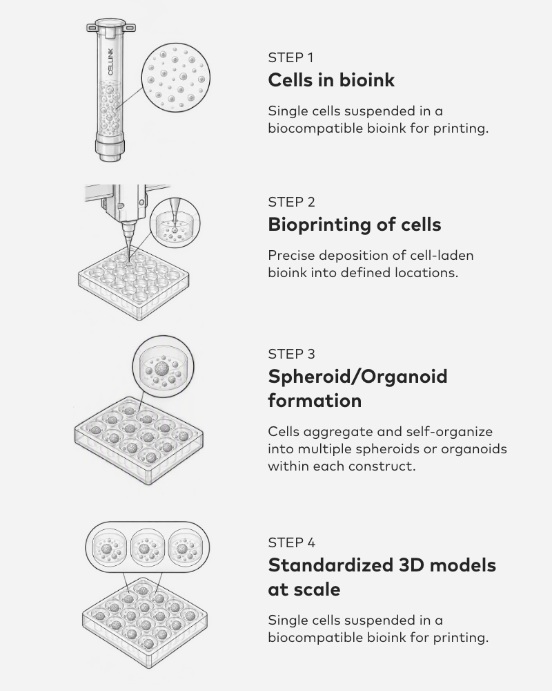
Single cells are suspended in a biocompatible bioink and deposited into defined locations using a bioprinter. Precise cell placement supports controlled aggregation and self-organization, allowing multiple spheroids or organoids to form within each printed construct. The workflow produces standardized 3D models with consistent size and distribution across wells, enabling scalable model generation for screening and research. The resulting constructs can be maintained in static or dynamic in vitro culture and adapted for tissue maturation or cell-delivery studies.
CELLINK solutions commonly used for this strategy include BIO ONE for standardized low-volume dispensing or BIO X, BIO X6 and BIONOVA X for scalable cell-laden bioprinting workflows.
What this enables
Automated Formation
Generate spheroids and organoids with minimal manual handling.
Reproducible Sizes and Controlled Placement
Consistent cell deposition supports reproducible model formation across wells and experiments.
Scalable Model Generation
Produce large numbers of models for screening and research.
Ready for In Vitro Dynamic Culture and In Vivo Cell Delivery
Suitable for in vitro culture, tissue maturation, and cell delivery studies.
Research examples
Regenerative tissue formation
Functional precision oncology
High-throughput cancer screening
Strategy 2: Print Spheroids or Organoids
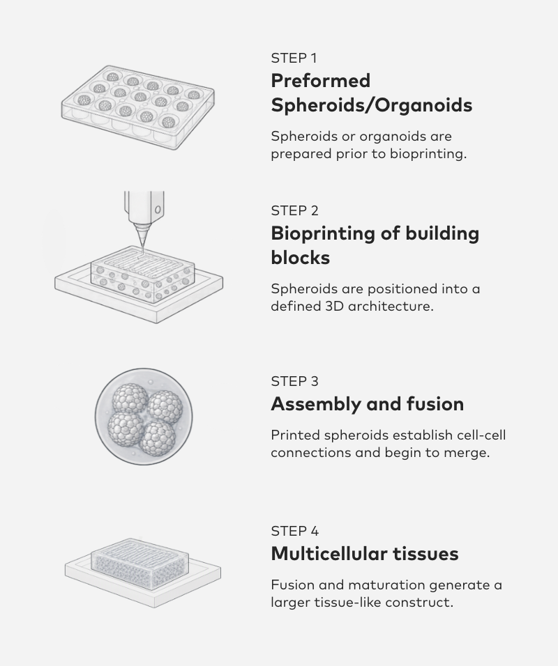
Preformed spheroids or organoids are used as biological building blocks, preserving the cellular organization and biological characteristics established before bioprinting. The building blocks are precisely positioned within a defined 3D architecture, where they establish cell–cell connections and begin to merge. Through continued fusion and maturation, the individual spheroids or organoids develop into a larger, multicellular tissue-like construct with increased structural and biological complexity.
This strategy can be implemented using CELLINK BIO X Series bioprinters and compatible biomaterials that support placement of spheroids and organoids within defined architectures.
What this enables
Preserve Established Biology
Preformed spheroids and organoids retain the cellular organization and biological characteristics developed before bioprinting.
Position with Precision
Bioprinting enables controlled placement of spheroids and organoids within defined spatial arrangements and 3D architectures.
Build More Complex Tissues
Multiple building blocks can be combined to create larger multicellular constructs with increased structural and functional complexity.
Research examples
Tumor microenvironment engineering
Disease modelling & drug discovery
Regenerative medicine & transplantation
Strategy 3: Print the Scaffold
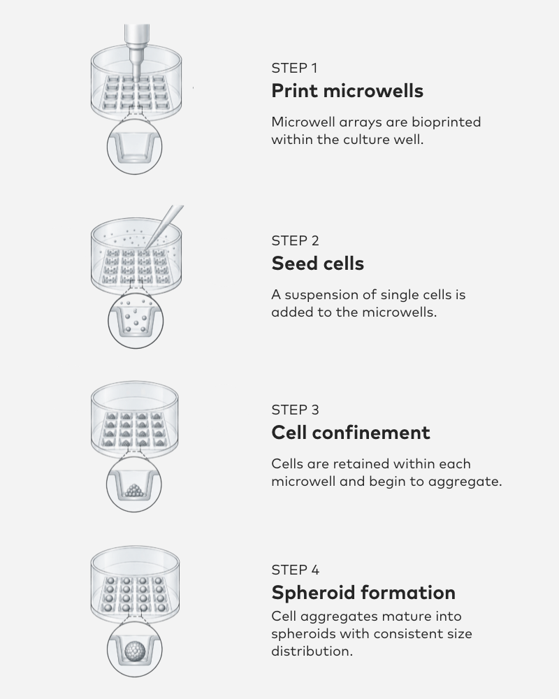
Bioprinting is used to create microwell arrays with defined geometry directly within the culture well. A single-cell suspension is then seeded into the microwells, where the printed structures confine the cells and promote controlled aggregation. Over time, the cell aggregates mature into spheroids with consistent size distribution. By combining standardized microwell geometry with a tunable material microenvironment, the workflow supports more reproducible spheroid formation across wells and experiments.
CELLINK LUMEN X and BIONOVA X bioprinters and biomaterials can be used to fabricate microwells, hydrogel compartments, and engineered culture environments that guide spheroid and organoid development.
What this enables
Defined Geometry
Bioprinted culture platforms provide control over well shape, spacing, and architecture, supporting consistent model formation.
Tunable Microenvironment
Material composition, mechanics, and biochemical cues can be adapted to influence cell behavior and organoid development.
Improved Reproducibility
Controlled culture conditions support more consistent spheroid and organoid formation across experiments and analytical workflows.
Research examples
Tissue engineering
Microenvironment engineering
Organoid patterning & development
Recent Publications

A 3D bioprinted cortical organoid platform for modeling human brain development
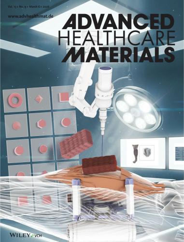
Highly Tunable and Cell-Remodelable Thiol-ene Alginate-Peptide Crosslinked Hydrogels to Recreate Cellular and Organoid Microenvironments for Biofabrication
Spatial engineering of posterior organizers in cerebral organoids via controlled morphogen exposure within hydrogels
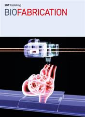
Cancer cell migration depends on adjacent ASC and adipose spheroids in a 3D bioprinted breast cancer model

3D bioprinting of liver spheroids derived from human induced pluripotent stem cells sustain liver function and viability in vitro