Applications / Disease Modeling
Disease Modeling
Model Human Disease with Greater Physiological Relevance
Disease modeling enables researchers to study human disease mechanisms, progression and therapeutic responses in controlled experimental systems. 3D disease models, such as bioprinted tissues, can better reproduce the complexity of human biology than traditional 2D cell cultures.
What is disease modeling?
In biomedical research, disease modeling refers to the practice of recreating biological pathologies in a controlled lab environment. The goal of disease model research is to study how diseases originate, advance, and respond to therapeutic interventions. By incorporating human cells, an extracellular matrix (ECM), and biophysical cues, modern 3D disease models reproduce complex cell-cell and cell-matrix interactions that traditional 2D cell cultures can’t match. These models provide physiologically relevant platforms to study disease initiation, track progression, and to accelerate preclinical drug discovery. Disease models are also used for personalized medicine through the creation of patient-specific models using the patient’s own cells.
Choosing the right model
The biological relevance of disease modeling depends on how closely a model reproduces the features of human tissue that matter to the research question. While 2D cell culture offer simplicity and scalability, 3D cell model approaches will capture additional features such as cell-cell interactions, tissue architecture, and extracellular matrix. 3D bioprinted models additionally offer greater control of the model design and cellular environment when compared to other 3D cell model methods, enabling greater reproducibility and more in vivo-like results.
Researchers therefore have to choose a disease model that provides the right balance of complexity, control, and throughput for their application.
Less Complex
More Complex
2D CELL CULTURE
Simple and scalable with limited representation of tissue complexity
2D cultures offer a controlled way to study cellular behavior and disease mechanisms. However, their flat environment cannot reproduce the 3D architecture, cell–cell interactions, and extracellular matrix of native tissue.
SPHEROIDS
3D models that capture cell-cell interactions and spatial organization
Spheroids bring cells together in 3D arrangements, enabling cell-cell interactions for cancer research, drug screening, and toxicity studies. Bioprinting enhances these models with uniform sizing, automated placement, and repeatable matrix encapsulation.
ORGANOIDS
Self-organizing models that reproduce selected features of human tissue biology
Organoids reproduce selected features of tissue organization and function to study disease mechanisms, development, and therapeutic responses. Bioprinting enhances these self-organizing models by adding precise spatial arrangement and batch-to-batch reproducibility.
BIOPRINTED TISSUES
Highly controlled 3D models that recreate tissue architecture and microenvironments
Bioprinted tissues can recreate specific tissue architectures, spartially organize multiple cell types, and incorporate ECM and biomaterials components. These models are useful for modeling complex tissues and disease-specific microenvironments.
HOW CELLINK CONTRIBUTES TO
Physiologically relevant disease models
Disease is often driven by more than a single cell type or biological pathway. It emerges from interactions between cells and the surrounding microenvironment, from the ECM to tissue architecture. 3D bioprinting enables researchers to adopt 3D cell culture models that recreate more of these factors in vitro, providing a controlled environment to investigate disease biology.
CELLINK 3D bioprinters and biomaterials support researchers in building 3D disease models with control over cell type, cell density, spatial organization, biomaterial composition, and mechanical properties. From spheroids to organoids and multicellular, tissue-specific constructs, these approaches bridge the gap between conventional 2D culture and the complexity of human tissue.
Researchers can print with diseased and healthy cells, or they can investigate how tissues change from physiological to pathological states by introducing disease-relevant conditions into healthy models. This enables studies of disease initiation and progression, as well as comparisons of therapeutic responses between healthy and disease tissue.
Disease Modeling Applications
HIGHLIGHTED RESEARCH & PUBLICATIONS
Cancer Models
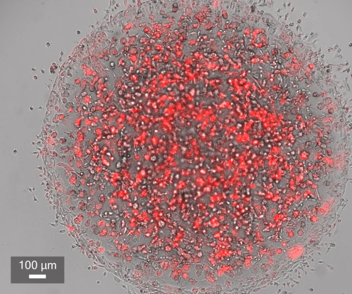
Cancer development is strongly influenced by interactions between tumor cells and their surrounding microenvironment. By combining multiple cell types with tunable biomaterials and defined tissue architectures, 3D bioprinted cancer models can recreate key aspects of tumor biology and provide a more physiologically relevant platform for studying disease progression and therapeutic response.
CELLINK bioprinting and biomaterials provide control over cell placement, biomaterial composition stiffness, and tissue architecture, supporting the development of complex, reproducible, and highly customizable cancer models.
Research examples
In Vitro Models for Leukaemia Studies
Bioprinted Head & Neck Tumor Models
Off-the-Shelf 3D Tumor Models
Organoids and Spheroid Models
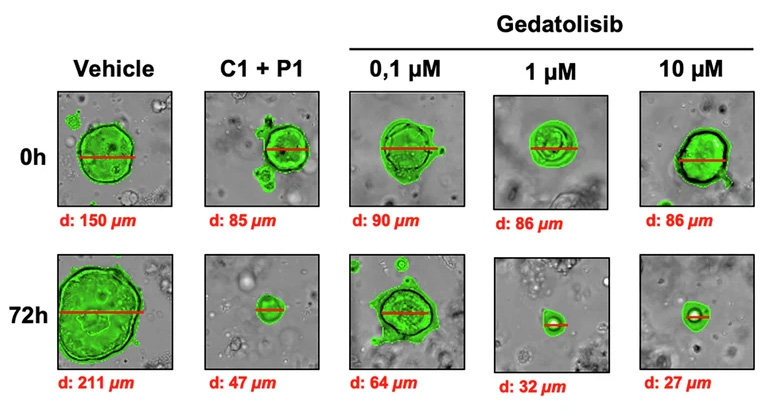
Organoids and spheroids are accessible 3D cell models for studying cell-cell interactions, tissue organization, disease phenotypes, and therapeutic responses. While spheroids offer relatively simple and scalable models, organoids can develop more complex, tissue-like structures and cellular organization.
There are three key strategies researchers use with CELLINK technologies when working with spheroids and organoids. Researchers can generate these models by depositing cells in a compatible bioink and allow them to aggregate and self-organize, place preformed biological units in a printable matrix and defined architecture or Create microwells, scaffolds or hydrogel compartments of choice before introducing the model. These approaches improve reproducibility, enable customized microenvironments, and support the development of advanced 3D models for research and drug discovery.
Research examples
Molecular Characterization of PARP Inhibitor Response
Bioprinted Organoid Microenvironments
3D Neurospheroid Brain Niche Models
Stem Cell Based Models
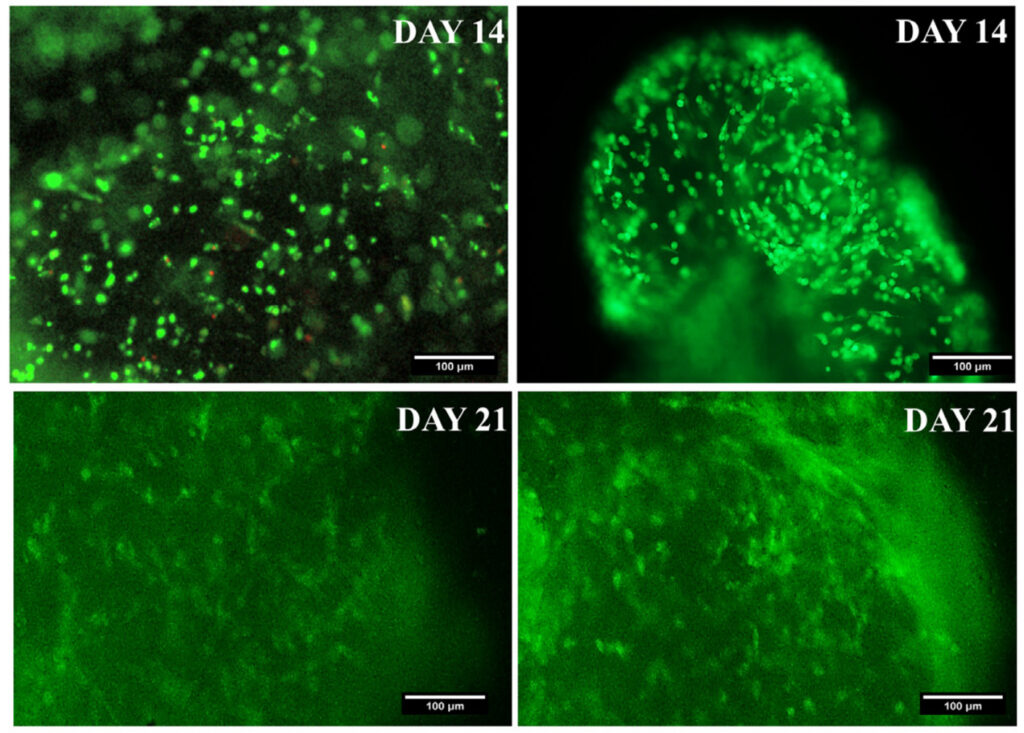
Stem cells are widely used to create physiologically relevant tissue models due to their ability to self-renew and differentiate into specialized cell types. Culturing stem cells within biomaterial-based 3D environments allows researchers to study differentiation, maturation, organization, and function within a tissue-like environment.
Developed stem cell-derived tissue constructs can be used to study disease progression and pathological changes by introducing pathogens or diseased cells to the construct.
Research examples
Bioprinted iPSC Heart Tube Models
Gradient-Guided Stem Cell Models
High-Density Stem Cell Bioprinting
Recent Publications

Molecular Characterization of PARP Inhibitor Response Reveals Co-Targeting Strategies in Advanced Prostate Cancer

Biphasic Granular Bioinks for Biofabrication of High Cell Density Constructs for Dermal Regeneration

Development of a 3D bioengineered human lung submucosal gland ductal airway model to study mucociliary clearance in vitro

AI-guided 3D bioprinting of multi-drug-loaded chitosan methacrylate hydrogels for personalized diabetic ulcer repair

Modeling early human heart development using an iPSC-based 3D bioprinted model of embryonic heart tube