A functional conductive bioink with physiologically relevant conductivity unlocks new possibilities for tissue modelling. It can enhance the transfer of electrical signals between neural cells, which is vital for neural tissue functionality and regeneration. Additionally, it can be used for the creation of muscular contraction models where cells are electrically stimulated to resemble a functional muscle.
The aim of the following project is to evaluate Bio Conductink based on GelMA supplemented with electrically conductive single-walled carbon nanotubes (CNTs).
Printing protocol
The printing protocol for Bio Conductink is optimized for the INKREDIBLE+ or BIO X equipped with our Temperature-controlled Printhead and a nozzle insulator. The bioink can be mixed with the cells in 10:1 ratio and printed with high precision, followed by quick and efficient photocrosslinking.
Electrical conductivity
Electrical conductivity facilitates communication between neural or muscle cells through electrochemical signals. Bio Conductink substantially increases the electrical conductivity of cell surroundings to 5·10-2 S/m, while retaining cell viability and functionality.
This video shows an LED lighting up as a part of an electrical circuit in the photocrosslinked conductive brain construct. The construct was bioprinted with the BIO X using Bio Conductink.
Viability
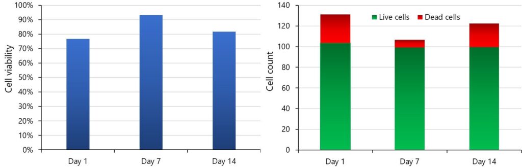
After printing, we assessed the cell viability of human dermal fibroblasts (HDFs) on days 1, 7 and 14. Despite the high stress cells encounter during bioprinting, HDFs exhibit a high viability of ≥70% on Day 1, which later rises to more than 80% (see left Figure beside). The presence of well-dispersed CNTs does not interfere with a common analytical method like fluorescence microscopy, which allows relatively easy detection of stained cells (see right Figure beside).
Experiment with neural cells
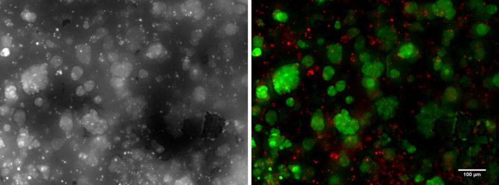
To show the effects of Bio Conductink on cells with electrical signalling ability, we performed a study with sensitive neural progenitor cells (NPCs). Due to their sensitivity, NPCs have somewhat higher number of dead cells (appear in red in the right Figure beside) in comparison to the HDF cell study presented above. However, our study proves Bio Conductink is compatible with neural cells and promotes formation of cell clusters (see bright-field Figure beside) that interact with each other through the hydrogel substrate.
Conclusion
This study illustrates great potential for CELLINK Bio Conductink in applications like high-precision printing and cell culturing. We successfully cultured two different cell types (HDFs and NPCs) in bioprinted constructs for 14 days and demonstrated the bioink’s universal biocompatibility.