Spheroids are three-dimensional (3D) cell cultures that self-aggregate into sphere-like formations during cell proliferation. While spheroids have been used within cell culture since the 1950s, it wasn’t until the early 1970s that the phenomenon was defined as a spheroid.
Traditional cell culture vs 3D spheroid cell culture
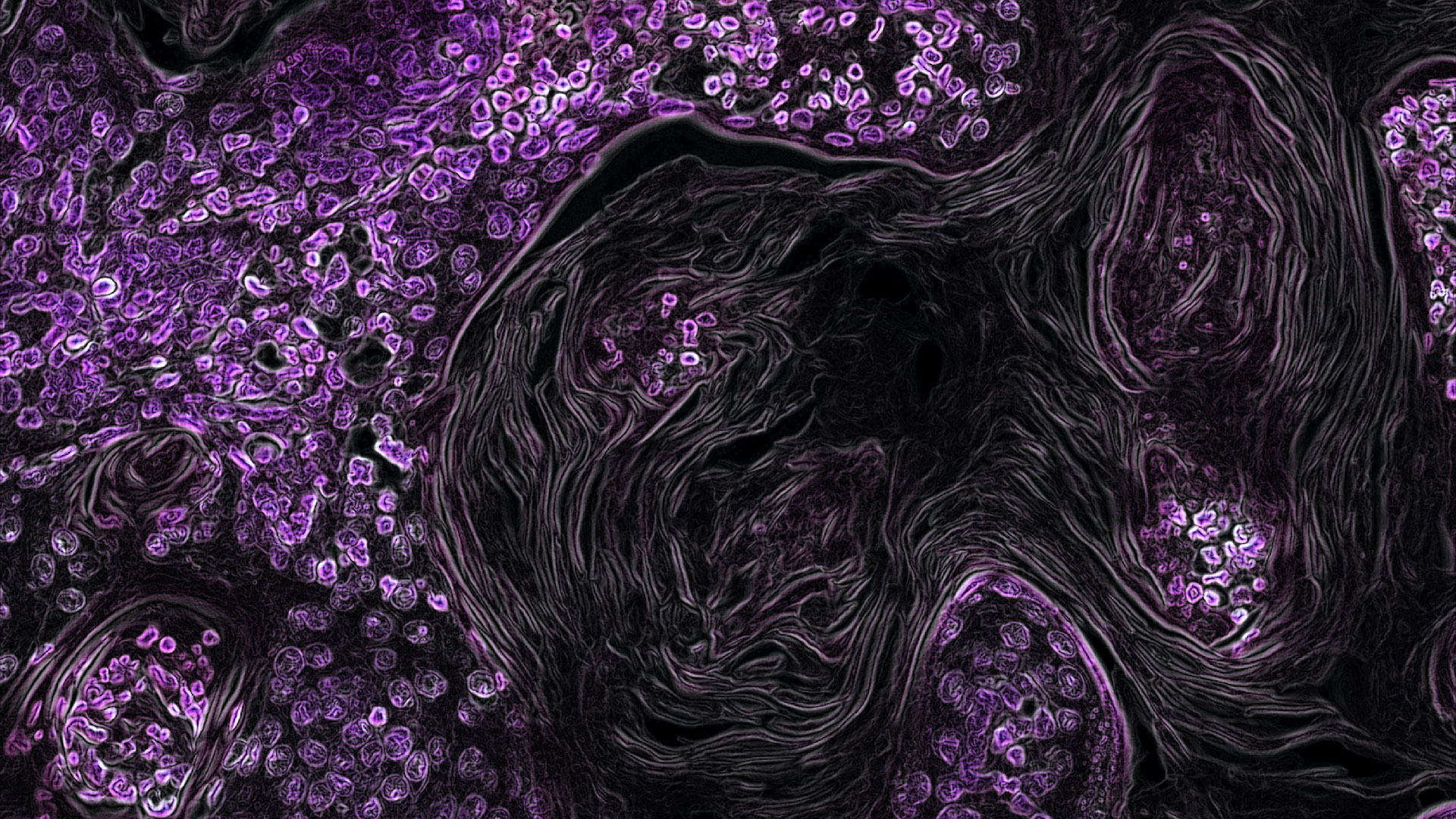
With two-dimensional, monolayer cell culture, cells primarily interact with the substrate they grow on. Meanwhile, 3D cell culturing can be used to produce spheroids which mimic the different cell interactions in native tissue, offering an insight into both diseased and normal tissues.
3D cell cultures also address the metabolic and functioning limitations that monolayer cultures face by mitigating the reduced cell-cell and cell-ECM interactions that limit cellular responsiveness.
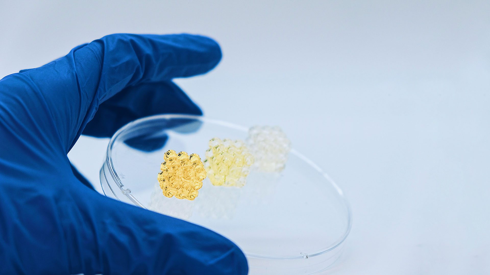
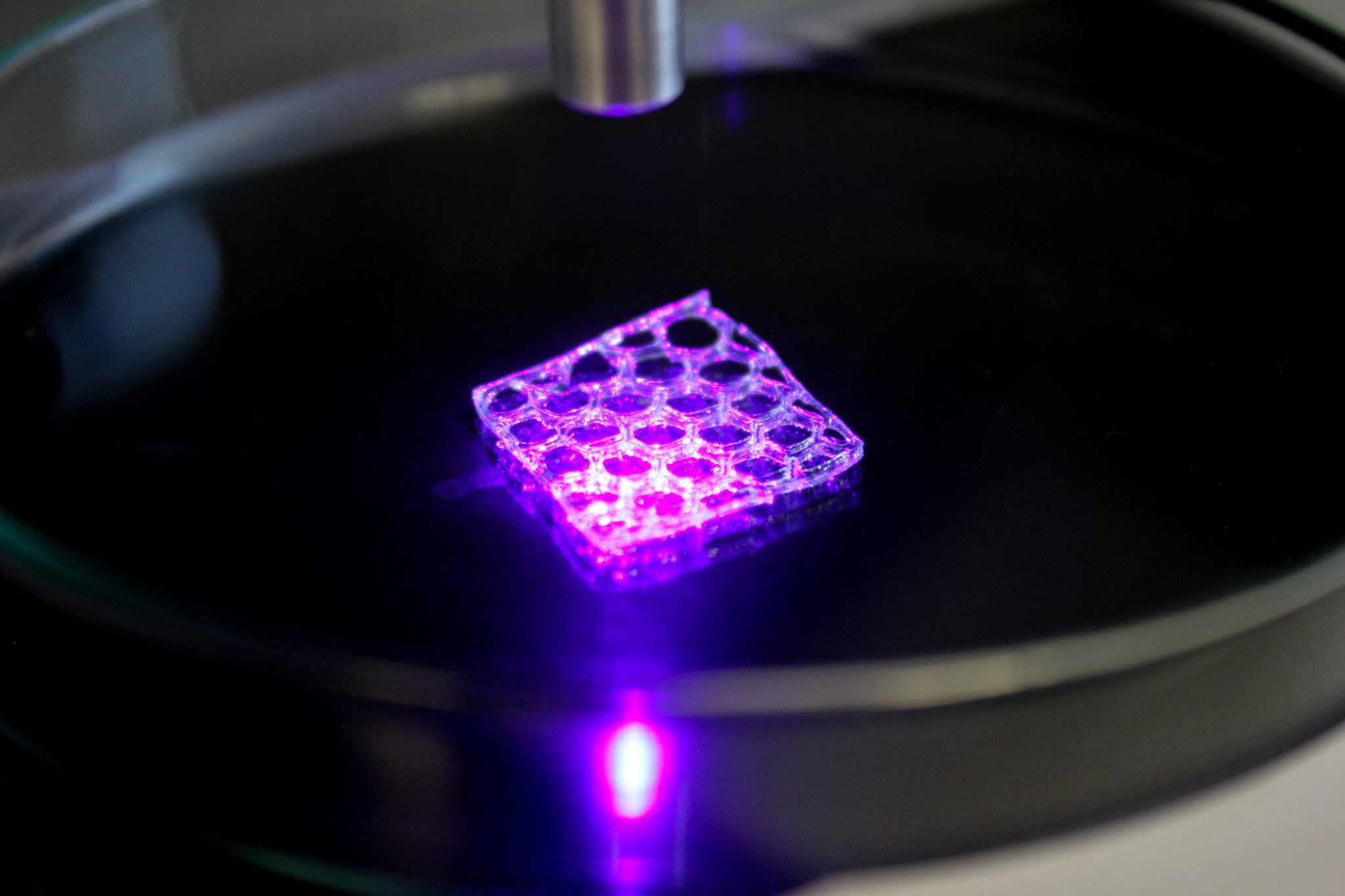
Depending on the cell line, spheroid formation can be further optimized within 3D cell culture by embedding them in a hydrogel or bioink, which mimics the cells’ ECM – the extracellular matrix. Embedding the spheroids in a bioink with relevant ECM components will promote both cell connectivity and tissue-like behavior, resulting in improved fidelity and resemblance to in vivo tissues.
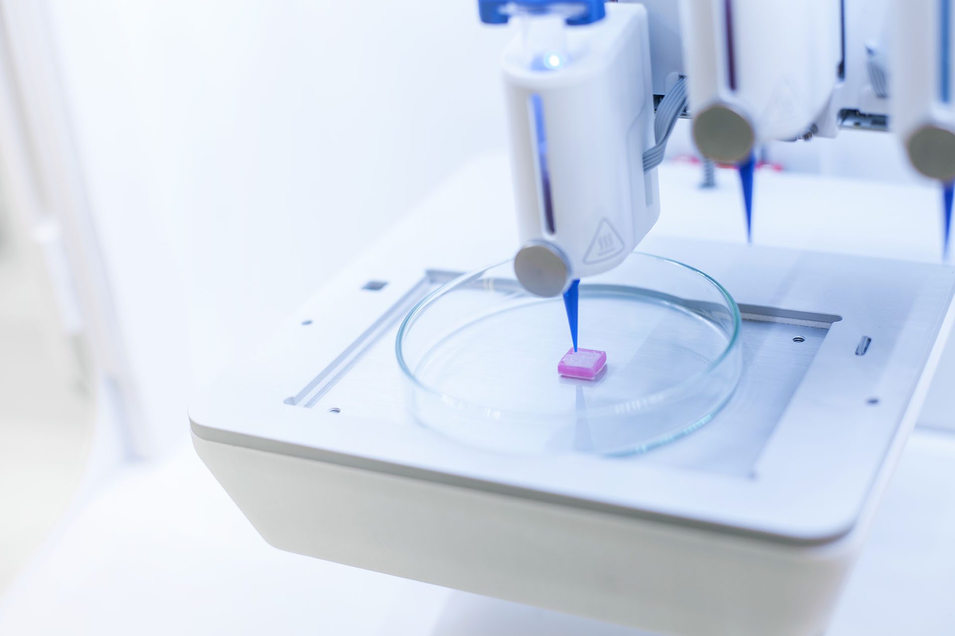
By using 3D bioprinting methods to produce constructs for spheroids, we can better understand cellular microenvironments. In turn, this means we can offer more physiologically relevant models for disease modelling and drug responsive mechanisms, eventually leading to drug discovery.
Spheroid applications
As mentioned in the introduction of this blog, spheroids are becoming increasingly important in understanding cancer behavior, improving drug screening, and enabling greater regenerative medicine. Below we talk a little bit more about how spheroids help, as well as list a few publications which utilize a bioprinting approach.
Tissue generation
Spheroids formed with mesenchymal stem cells (MSCs) have shown great tissue regeneration and repair properties. This is no surprise, as MSCs are multipotent cells that have regenerative properties. The usage of cell clusters of MSCs has the potential to increase survival periods after tissue implantation.
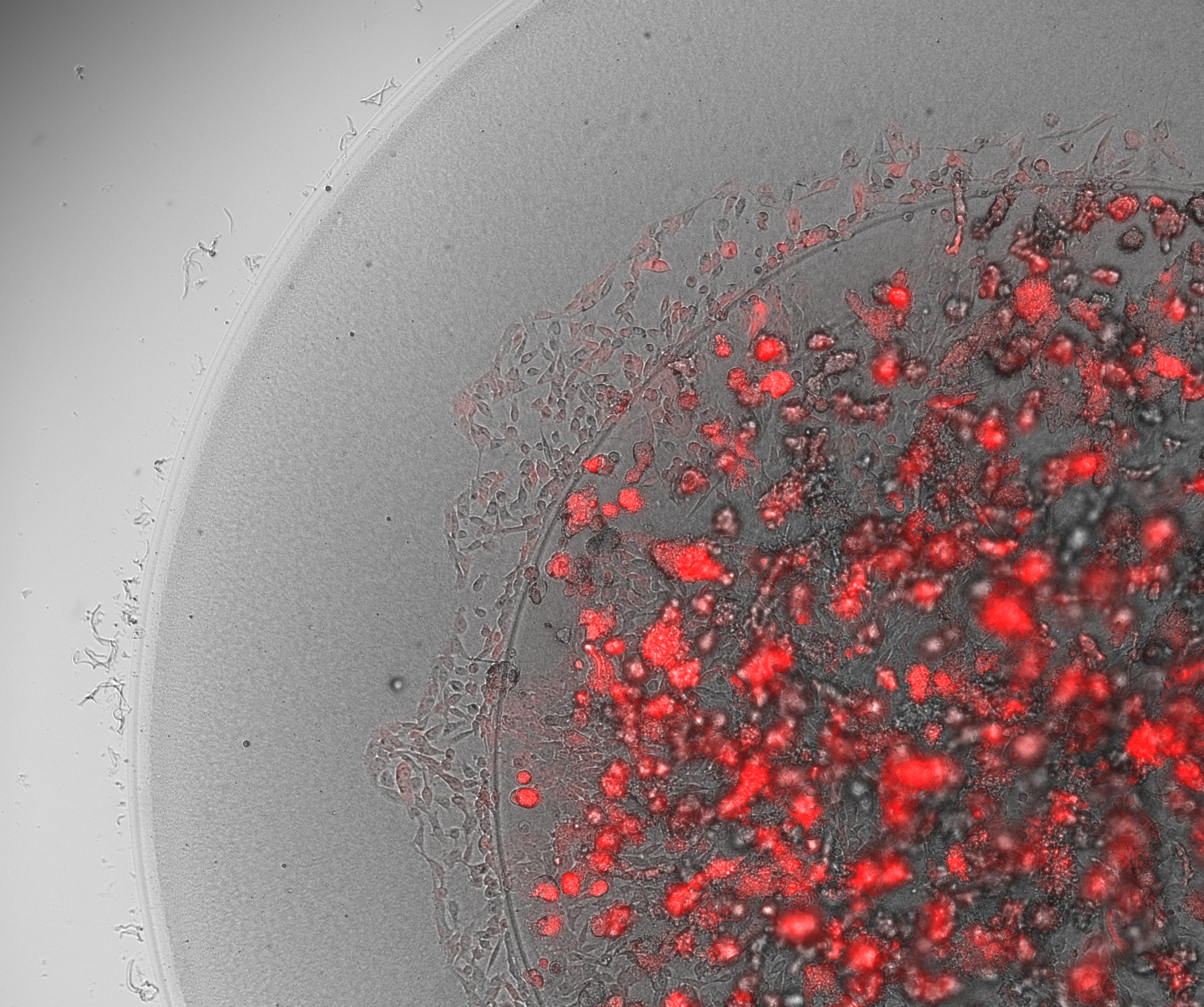
The use of tumor spheroids within cancer research is becoming increasingly common. A tumor spheroid is an in vitro spherical 3D cell assembly of tumor cells. These are designed to mimic aspects of the in vivo tumor microenvironment, and they are used for various purposes: drug screening, understanding tumor biology, and investigating cancer therapies.
Within cancer research, researchers often use multicellular tumor spheroid models, MCTS. The unique cell composition within an MCTS reveals how cells grow, interact, proliferate, and absorb nutrients. These properties make spheroids ideal pre-clinical test models for drug discovery.
Becconi, De Zio, Falciani et al. (2023) Cancers 15(4)
3D bioprinting to automate spheroid formation
While spheroids are continuing to be used within research, there have traditionally been challenges with finding suitable techniques for the formation of spheroids, which combine increased throughput, reproducibility and size uniformity. The latter is especially important for physiologically relevant research results, as cellular functions inside spheroids show a correlation to the size of the cell cluster.
With 3D bioprinting technologies, we are accelerating spheroid formation for better predictive tissue models. These technologies enable improved spatial control, reduce human error, offer great material versatility and faster speeds. Scientists can develop standardized workflows, avoid time-consuming and repetitive tasks, and gather more consistent data.
Create your own
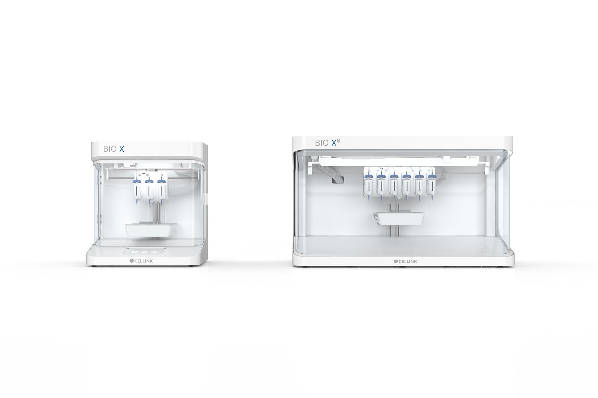
At CELLINK, we recognize the need for tools that simplify the reproducible formation of these cell clusters.
Our bioprinters and biodispensers promote formation of spheroids by allowing for multi-material printing of various biomaterials, including hydrogels, matrix components, and other key biologically relevant components. These instruments, both bioprinters and biodispensers, support any* cell type and offer high-precision droplet dispensing. We believe that refining the efficiency of spheroid formation with scalable and cost-efficient 3D bioprinting systems will promote the study of cellular biology. Advances in this field will no doubt lead to new and exciting biomedical applications.
Application notes
High-throughput Spheroid Formation and Migration with TeloCol-10 for Improved Stability
Demonstrating the use of Advanced BioMatrix’s TeloCol-10 for improved stability over longer incubation times compared to Matrigel, when bioprinting spheroids.
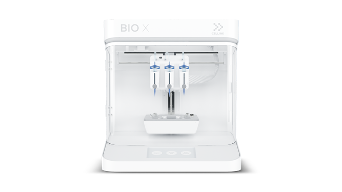
In Vitro 3D Lung Cancer Model Presents a More Relevant Expression of Junctional Proteins than 2D Cultures
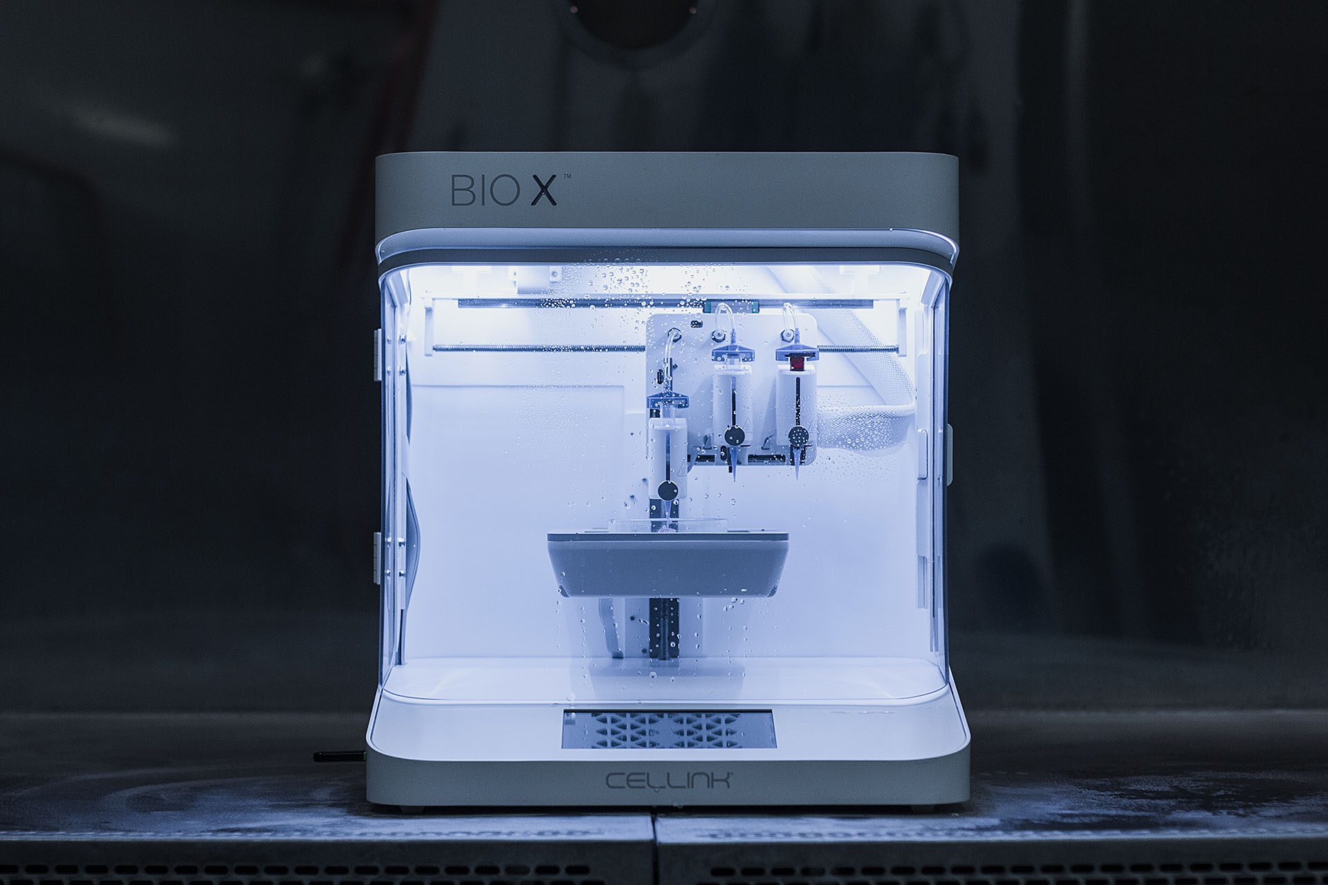
We compare human lung cancer cultured on 2D polystyrene plates compared to ones that were 3D bioprinted with the BIO X. In the 3D cell culture, there was a cellular rearrangement into spheroids and a distribution of junctional proteins which could not be detected in the 2D cultures.
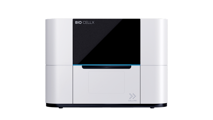
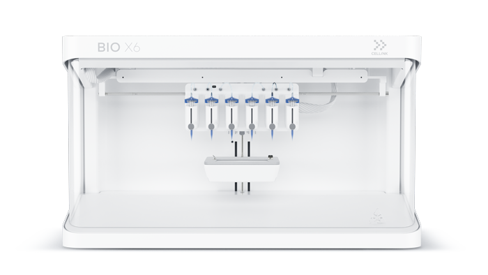
Biodispensers & Bioprinters for spheroid formation
CELLINK is leading the way in 3D bioprinting and biodispensing instruments, with more than 1200 publications using our bioprinters across the spectrum of life science applications areas.